


More Related Content
Similar to Urolo radiology eng procedure by Van Scri (20)
More from VAN DINH (19)


Recently uploaded (20)


Urolo radiology eng procedure by Van Scri
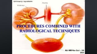
- 1. PROCEDURES COMBINED WITH RADIOLOGICAL TECHNIQUES Au.: MD Van Scri ŌĆō VN 2023
- 2. OUTLINE USUALLY COMBINES 3 AREAS: - CLINICAL. - SUBCLINICAL. - IMAGE ANALYSATION + ULTRASOUND. + UIV. +UPR. + UCP. +CT. + RENAL ANGIOGRAPHY.
- 3. SUPERSONIC DIAGNOSTIC TOOLS ARE VERY COMMON. OBSERVE THE KIDNEYS, RENAL PELVIS, AND KIDNEYS. RENAL FUNCTION COULD NOT BE ASSESSED. 1. TECHNICAL. - PATIENTS DO NOT NEED TO PREPARE. - PATIENT HOLDS URINE IF POSSIBLE. - USEFUL IN CHILDREN AND UNCOOPERATIVE PATIENTS. - PORTABLE SA CAN BE USED AT BED.
- 4. SUPERSONIC 2. INDICATION. - IN UROLOGICAL DISEASES. + KIDNEY CYSTS, KIDNEY TUMORS. + KIDNEY HYDRONEPHROSIS, KIDNEY ASSESSMENT. - URINARY STONES. - OF LITTLE VALUE: RENAL CALYX-PYELOPATHY, PERIRENAL TISSUE, ADRENAL, KIDNEY DISEASE, IN TRAUMA.
- 5. SUPERSONIC A. IN KIDNEY TRANSPLANT. - IS AN EXCEPTION. - USE SA DOPPLER TO OBSERVE: PELVIC ARTERY, RENAL ARTERY, INTERLOBAR ARTERY. - INDICATES WHETHER THE TRANSPLANTED KIDNEY HAS FAILED OR BEEN REJECTED.
- 6. SUPERSONIC B. SCROTUM, PENIS. - TESTICLES: INFLAMMATION, TUMORS, HYDROCELE. CYSTS, TUMORS, INFLAMMATION, EPIDIDYMIS. - ASSESS DV: TUNICA ALBUGINEA, CORPUS CAVERNOSUM, CORPUS SPONGIOSUM, DV BLOOD VESSELS. - DILATES SPERM VEINS.
- 7. SUPERSONIC 3. THROUGH THE RECTUM. A. POINT. - INCREASED PSA, TUMORS, PROSTATE INFLAMMATION, OBSERVATION OF SEMINAL VESICLES, EJACULATORY DUCTS. - BIOPSY AND DRAINAGE OF PROSTATE ABSCESS B. TECHNIQUE. - RINSE THE INTESTINES AND URINATE BEFORE SA. KS IF ST. - T TILT OR OBSTETRICS AND GYNECOLOGY. - ST 6 SAMPLES WITH 18 GAUGE NEEDLE.
- 8. SUPERSONIC Normal kidney SA image
- 9. SUPERSONIC Images of SA BQ Normal BQ citadel is ruined
- 11. UIV (IVU) 1. TECHNICAL. - PATIENTS FAST FOR AT LEAST 6 HOURS BEFORE THE SCAN. - CLEANSES THE COLON. - COMPLETELY URINATE BEFORE TAKING THE SCAN. THE PATIENT LIES ON HIS BACK. - BLOOD UREA < 0.8G/L. - HOW TO MAKE A UIV MOVIE. + KUB EXAMINES THE ABDOMEN. + PRESS YOUR ABDOMEN WHEN TAKING THE PHOTO.
- 12. UIV (IVU) 2. INDICATION. - CHECK RENAL PELVIS, NQ, SUSPECTED UROTHELIAL TUMOR. SUSPECTED CONGENITAL ABNORMALITY OF THE URINARY SYSTEM. - AFTER BT-NQ AND NQ-BQ JUNCTION SURGERY. - KIDNEY STONES, NQ, DISTENDED KIDNEYS. - URINARY TRACT OBSTRUCTION. - TRAUMA, HEMATURIA, KIDNEY DISEASE,... - SUSPECTED COMPLICATIONS AND URINARY COMPLICATIONS AFTER SURGERY.
- 13. UIV (IVU) 3. CONTRAST AGENT. OFTEN USE DIODON, VIOSTRAST, CARDIOSTRAST... - HAS LOW PERMEABILITY. - HAS HIGH PERMEABILITY. - SINGLE OR MULTIMOLECULAR GROUPS THAT CREATE IONS: TEL├ēBRIX- 35, HEXABRIX-32... - NON-IONIC RADIOPAQUE GROUP: TRIENETIX-30...
- 14. UIV (IVU) 4. RESULTS. A. NORMAL. B. PATHOLOGICAL. - RENAL FUNCTION - BODY DEFORMITY. - BLOCKAGE LOCATION. - BLADDER. Normal UIV 30 minutes
- 15. UPR 1. INDICATION. - KIDNEY UIV DOES NOT ABSORB DRUGS, LEAVING THE HEART THE CAUSE OF OBSTRUCTION. - FIND FISTULA ROUTES THROUGH THE LYMPHATIC SYSTEM. - RARELY USED DUE TO PAIN AND UPSTREAM INFECTION. - SPECIFY ONLY WHEN ABSOLUTELY NECESSARY.
- 16. UPR 2. PROCEDURES. INJECT 12 ŌĆō 15 ML OF CONTRAST MEDIUM. MOVIE 1 AFTER INJECTION, MOVIE 2 AFTER 5 MINUTES. 3 RESULTS. -RENAL PELVIS, NQ LIKE UIV. -DETERMINE THE LOCATION OF FOREIGN BODIES, STENOSIS IN THE KIDNEY OR NQ. -FISTULA FROM KIDNEY TO LYMPHATIC SYSTEM.
- 17. RETROGRADE CYSTOGRAPHY. 1. INDICATION. INJURY, PROLAPSE, URINARY TRACT FISTULA. POSTOPERATIVE. BQ BODY SHAPE. EVALUATE URINE LEAKAGE. 2. TECHNICAL. SOFT CATHETER. ADEQUATE CONTRAST AGENT. 3. RESULTS. - BQ -NQ REFLUX. BQ -INTESTINAL PROBE. - PROBE THE BQ -UTERUS INTO THE AD. U BQ, TLT. - DILATION, INFLAMMATION, PROLAPSE OF THE BLADDER, BLADDER NERVES.
- 18. RETROGRADE CYSTOGRAPHY. BQ normal Tune BQ ŌĆō TC into AD
- 19. RETROGRADE URETHROGRAPHY. 1. INDICATION. 2. TECHNICAL. INJECT THE MEDICINE UPSTREAM FROM THE MOUTH OF THE FLUTE. 3. RESULTS. - NORMAL. - PATHOLOGICAL. + ND STENOSIS. NDŌĆōTSM, NDŌĆōRECTAL FISTULA. + DEFORMATION OF THE URETHRA.
- 21. CT URINARY SYSTEM 1. HISTORY. - INVENTED BY ENGINEER GODFREY NEWBOLD HOUNSFIELD AND HIS COLLEAGUES. - 1971 THE FIRST BRAIN CT WAS BORN, CUTTING ONE LAYER TOOK 4 MINUTES. - DEVELOPED THROUGH 4 GENERATIONS.
- 22. CT URINARY SYSTEM 2. OPERATING PRINCIPLE. - THE MOVING X-RAY SOURCE SCANS CROSS-SECTIONAL LAYERS AT DIFFERENT DEGREES. - BASED ON THE DENSITY OF EACH BODY PART, DIFFERENT IMAGES ARE PRODUCED. - DIAGNOSIS BASED ON DENSITY: WHITE (BONE), BLACK (FLUID, WATER, VAPOR).
- 23. CT URINARY SYSTEM 2. OPERATING PRINCIPLE. - HOUNSFIELD UNIT (HU) TO MEASURE DENSITY: + WATER IS 0HU. + GAS IS 1000HU. - THERE ARE 3 LEVELS OF DENSITY: + CONCENTRATED COPPER. + INCREASE DENSITY. + REDUCE DENSITY.
- 24. CT URINARY SYSTEM 3. TECHNICAL. - CUT THE LAYER ALONG THE CONVENTIONAL AXIS, THE SCANNING TABLE SLIDES STEP BY STEP THROUGH THE SCANNER. - SPIRAL CT IS MORE ACCURATE THAN CONVENTIONAL CT. + CONTINUOUS SLIDING TABLE. THE PATIENT HELD HIS BREATH ONCE. + PITCH = TABLE SLIDING SPEED / LAMP OPENING = 1:1, TAKING 1 KIDNEY SHOT TAKES 30 SECONDS.
- 25. CT URINARY SYSTEM 3. TECHNICAL. - SPIRAL CT DOES NOT HAVE DEVIATIONS DUE TO MOVEMENT AND SPACE LIKE CONVENTIONAL CT. - CT WITH FLUOROSCOPY. + THE PATIENT FASTED FOR 4 HOURS BEFORE INJECTING THE DRUG. + INJECT 100ML OF MEDICINE, 1.5 - 4 ML/S. - THERE ARE MANY TYPES OF DRUGS ON THE MARKET.
- 26. CT URINARY SYSTEM 3. TECHNICAL. STAGES AFTER INJECTION: - MM PHASE: AFTER 15 - 40 SECONDS. - MARTIAL KIDNEY STAGE: AFTER 25 - 80 SECONDS. - RENAL PHASE: AFTER 90 - 120 SECONDS. - EXCRETION PHASE: AFTER 3 - 5 MINUTES.
- 27. CT URINARY SYSTEM 4. HOW TO DO IT. A. KIDNEY STONES, NQ. B. KIDNEY TUMOR. C. RENAL BLOOD VESSELS. D. URINARY TRACT INFECTION. E. BQ AND NQ.
- 28. CT URINARY SYSTEM Renal blood vessels
- 29. RENAL ARGIOGRAPHY 1. INDICATION. - HEMATURIA SUSPECTED OF VASCULAR ABNORMALITIES. - KIDNEY TUMOR: VASCULAR DISTRIBUTION. - BEFORE SURGERY: PARTIAL NEPHRECTOMY, LARGE KIDNEY, ADRENAL, RETROPERITONEAL TUMORS. - RENAL VASCULAR DISEASE. - SUSPECTED RENAL VASCULAR INJURY ON UIV OR CT IN TRAUMA.
- 30. RENAL ARGIOGRAPHY 2. PRINCIPLES. - TAKING 2 RENAL ARTERIES TOGETHER: INJECTING MEDICINE INTO THE AORTA ABOVE THE RENAL ARTERY. - SCAN EACH RENAL ARTERY SEPARATELY: FROM THE AORTA, INSERT THE CATHETER INTO THE RENAL ARTERY TO BE SCANNED AND INJECT MEDICATION.
- 31. RENAL ARGIOGRAPHY 3. PROCEED. A. DIRECT METHOD. - INSERT THE NEEDLE DIRECTLY INTO THE AORTA ABOVE THE RENAL ARTERY. B. INDIRECT METHOD. - CATHETER FROM THE FEMORAL ARTERY UP TO THE 12TH LUMBAR AND 1ST LUMBAR VERTEBRAE. MEDICATION PUMP. - DIRECT THE CATHETER INTO THE KIDNEY TO TAKE THE SCAN. KIDNEY SCAN TO CHOOSE FORTUNE.
- 32. RENAL ARGIOGRAPHY 4.RESULTS. A. NORMAL. CLEARLY SEE THE DIVISION OF MM INTO EACH KIDNEY. B. PATHOLOGICAL. - MM PROLIFERATION IN MALIGNANT TUMOR AREAS. - REDUCE MM IN KIDNEY CYST AREA. - RENAL ARTERY ANEURYSM. - RENAL ARTERY STENOSIS.
- 33. RENAL ARGIOGRAPHY 5. COMPLICATIONS. - THROMBOSIS. - MM PSEUDOANEURYSM. - ARTERIAL EMBOLISM. - DISSECTION INTO MM. - ALLERGY OR NEPHROTOXICITY DUE TO CONTRAST DYE.
- 34. COMPARE THE VALUES OF DIAGNOSTIC IMAGING METHODS Kidney tissue owner Kidney stones Renal function Renal pelvis Ureters Bladder KUB + + + 0 0 0 + UIV + + + + + + + + + + + + + + + Capture upstream 0 + + + + + + + + + + + + + Supersonic + + + + + + 0 + + + 0 (if not stretched) + + (if stretched) Clearly seen through cystoscopy CT without contrast + + + + + 0 + + + + + + + CT with contrast + + + + + + + + + + + + + + + + + + + MRI + + + + 0 + + + + + + + + + + + Kidney scintigraphy + + 0 + + + + + + + + + +
- 35. A. KIDNEY AND URETER STONES. - REPLACE UIV IN RENAL COLIC. - CONTRAST-ENHANCED CT SHOULD NOT BE USED TO MISDIAGNOSE INTESTINAL DIVERTICULA. - CT USUALLY DOES NOT SHOW STONES OR WHEN IT IS NECESSARY TO DETERMINE KIDNEY FUNCTION, A SLOW CONTRAST CT SCAN IS PERFORMED AFTER 10 MINUTES.
- 36. B. KIDNEY TUMOR. -GET A PLAIN CT SCAN FIRST. -ONE FILM 1 MINUTE AFTER DRUG INJECTION. -AFTER 10 MINUTES, TAKE A FILM. MANY KIDNEY TUMORS CLEARLY SHOW THE EXCRETION STAGE. - SPIRAL CT IS DONE QUICKLY, SCANS CONTINUOUSLY, AND ALWAYS MEASURES BLOOD VESSELS. - SEE KIDNEY TUMOR INVADING VEINS, NUMBER OF ARTERIES.
- 37. C. RENAL BLOOD VESSELS. - IDENTIFY RENAL MM PATHOLOGY. - INJECT THE DRUG INTO THE ANTERIOR TIBIAL VEIN AT 3ML/S. - TAKE A PHOTO AFTER 20 - 25 SECONDS. SLOW FILM CLEARLY SHOWS THE STRUCTURE OF KIDNEY MM. - 2 OR 3 DIMENSIONAL IMAGING CLEARLY SHOWS MM ABNORMALITIES.
- 38. D. URINARY TRACT INFECTION. - USUALLY RELIES ON LS. CT TO DETECT COMPLICATIONS OR MONITOR TREATMENT. - CT OFTEN SHOWS ABNORMAL KIDNEYS. - CONTRAST-ENHANCED CT CLEARLY SHOWS THE LESIONS, WHEREAS CT SHOWS NO LESIONS. - NO SIGNS IN URINARY TRACT INFECTION.
- 39. E. BQ AND NQ. - TAKE A SHOT AFTER PUMP MEDICINE 5 - 10 MINUTES . LIE YES , YES CAN CONCLUDE VALSALVA MATCH . - TWISTED CT SNAIL SEE OCCLUSION BLOCKAGE AND INFLAMMATION INFECTED NQ PULSE . - SA PRICE TREAT THAN IN DAMAGE LOVE BQ. - CT SEES IT CLEARLY TISSUE FAT PULSE AROUND AND LYMPH NODES REGION POT .
- 40. A. KIDNEY AND URETER STONES. - REPLACE UIV IN RENAL COLIC. - CONTRAST-ENHANCED CT SHOULD NOT BE USED TO MISDIAGNOSE INTESTINAL DIVERTICULA. - CT USUALLY DOES NOT SHOW STONES OR WHEN IT IS NECESSARY TO DETERMINE KIDNEY FUNCTION, A SLOW CONTRAST CT SCAN IS PERFORMED AFTER 10 MINUTES.