Inflammatory Localized and Generalized Bone Loss in Recent-onset Rheumatoid Arthritis, Presentatie van dr. Melek Guler-Yuksel op 18 april 2012 voor de Stichting IWO.
The ASBMR 2011 conference presented numerous findings on osteoporosis and fracture risk, highlighting trends in circulating biomarkers, vitamin D, and calcium, among others. Key studies detailed the influence of inflammatory biomarkers on fracture risk and the effectiveness of bisphosphonates and denosumab in fracture reduction. Future directions stress the importance of including male participants in clinical trials and refining treatment guidelines for osteoporosis.
The document summarizes results from a phase 3 clinical trial of the drug odanacatib for treating osteoporosis in postmenopausal women. Key findings include:
- Odanacatib significantly reduced the risk of morphometric vertebral fractures by 54%, hip fractures by 47%, and nonvertebral fractures by 23% compared to placebo.
- Bone mineral density increased substantially with odanacatib therapy over 5 years.
- Safety analyses found low rates of adverse events with odanacatib, though some rare skin conditions were more common compared to placebo and require further investigation.
Dove no va il sole, va il medico en andere tegeltjes wijsheden (over vitamine D), Presentatie van dr. A. van de Wiel op 18 april 2012 voor de Stichting IWO.
The document discusses the relationship between obesity and fractures, highlighting that while obesity may protect against certain types of fractures in older women, it can also lead to increased fracture risk due to factors like mechanical instability and site-specific bone loss. It presents research findings, including the effects of fat and bone interactions, the role of vitamin D, and the impact of weight loss on bone density. Conclusions suggest obesity poses a complex risk factor for fractures, differing across age groups and health contexts.
The document discusses the mechanisms and clinical aspects of stopping denosumab and romosozumab treatments, highlighting accelerated bone loss after discontinuation. Key factors influencing bone density changes include baseline serum levels, treatment duration, and alternative therapies. The document also examines potential strategies to mitigate post-treatment bone loss, such as gradual dose reduction and the use of sclerostin antibodies.
1) DXA scanning is a reliable and low-radiation method to measure bone mineral density (BMD) at the lumbar spine, hip, and wrist to diagnose osteoporosis.
2) DXA can also detect vertebral fractures (VFA) and measure whole body composition, abdominal fat, and aortic calcification.
3) Interpretation of DXA results requires attention to potential variability between devices, accurate placement of regions of interest, and use of appropriate reference data since BMD can be under or overestimated in certain patients.
This document summarizes osteonecrosis of the jaw (ONJ) associated with antiresorptive agents. It defines ONJ and stages its severity. It discusses the pathogenesis of ONJ and risk factors like underlying disease, treatment duration, and dental procedures. Cancer patients on intravenous bisphosphonates have the highest ONJ risk of 1-8% due to higher drug doses and worse oral/general health. Management involves conservative measures like mouthwashes for early stages and surgery with antibiotics for later stages. Discontinuing antiresorptives may help healing but risks fractures. Teriparatide may help healing in some cases but its use in cancer is uncertain. More research is needed on preventing and treating established ON
This systematic review analyzed 895 cases of tumor-induced osteomalacia (TIO) from case reports. TIO is caused by tumors that produce excess fibroblast growth factor 23 (FGF23), which causes hypophosphatemia and osteomalacia. The review found that TIO mostly affects adults aged 40-60 years old, with long diagnostic delays of several years on average. The tumors were located variably but most commonly in the lower limbs or head and neck region. Higher FGF23 levels correlated with larger tumor size. Patients experienced significant bone fragility and fracture rates as high as 60% due to long-term hypophosphatemia. Early tumor detection and removal are important to improve outcomes for
This document discusses real-world evidence on denosumab for osteoporosis treatment and fracture prevention. It summarizes several studies, including one that found denosumab reduced fracture risk by 38% compared to placebo in over 25,000 postmenopausal women. Another study showed good long-term persistence with denosumab therapy in over 800 patients. Additional studies observed that zoledronic acid can prevent bone loss following denosumab discontinuation, and bisphosphonate treatment after denosumab provides protection against new vertebral fractures.
A nationwide registry study in the Netherlands reveals that patients with osteogenesis imperfecta (OI) have a hospitalization rate almost three times higher than the general population, primarily due to both skeletal and extraskeletal complications. The study involved 674 OI patients with 2,225 hospital admissions, indicating a mean length of stay of 4.5 days per admission. Increased awareness of OI and its impact on patients' lives is essential for improving prevention and follow-up care.
The document appears to be a presentation on highlights from the ASBMR 2021 conference in San Diego. It discusses several topics that were covered at the conference, including fracture risk assessment, the effects of various osteoporosis treatments on bone mineral density, safety issues like osteonecrosis of the jaw and atypical femoral fractures, the role of vitamin D, and applications of artificial intelligence. The entire document is copyrighted by Prof. Dr. Joop van den Bergh.
This document discusses guidelines for medication to prevent fractures in patients using glucocorticoids. It notes that glucocorticoids significantly increase the risk of vertebral and non-vertebral fractures. While effective anti-osteoporosis drugs are available, many glucocorticoid-treated patients remain untreated. The document reviews new guidelines that simplify treatment criteria to improve implementation and outlines recommendations for when to start bone-sparing medications based on patient factors and glucocorticoid dose and duration. The goal is to optimize fracture prevention in glucocorticoid-treated patients.
This document discusses what actions should be taken when a vertebral fracture is discovered incidentally. It notes that vertebral fractures are very common fractures, especially in older individuals, and are often asymptomatic. Having a vertebral fracture significantly increases one's risk for future fractures both in the short and long term. If a vertebral fracture is found incidentally, such as on a CT scan, further investigation is warranted including assessing bone mineral density and checking for underlying bone diseases. Treatment options should also be considered, especially if the individual has low bone density in addition to the vertebral fracture, as this combination confers the highest risk. New automated detection algorithms aim to help identify vertebral fractures on scans to ensure appropriate follow up for individuals.
This document summarizes a cost-effectiveness model of Fracture Liaison Services (FLS) care in the Netherlands. The model found that FLS care would be highly cost-effective, with a cost of 9,076 per quality-adjusted life year gained. Total 5-year costs with FLS would be only 1.7% higher than current costs but would prevent fractures and improve health outcomes. The model can help decision-makers prioritize secondary fracture prevention and allow local payers and FLS to predict costs and benefits of implementation.
More Related Content
More from Stichting Interdisciplinaire Werkgroep Osteoporose (20)
The document discusses the mechanisms and clinical aspects of stopping denosumab and romosozumab treatments, highlighting accelerated bone loss after discontinuation. Key factors influencing bone density changes include baseline serum levels, treatment duration, and alternative therapies. The document also examines potential strategies to mitigate post-treatment bone loss, such as gradual dose reduction and the use of sclerostin antibodies.
1) DXA scanning is a reliable and low-radiation method to measure bone mineral density (BMD) at the lumbar spine, hip, and wrist to diagnose osteoporosis.
2) DXA can also detect vertebral fractures (VFA) and measure whole body composition, abdominal fat, and aortic calcification.
3) Interpretation of DXA results requires attention to potential variability between devices, accurate placement of regions of interest, and use of appropriate reference data since BMD can be under or overestimated in certain patients.
This document summarizes osteonecrosis of the jaw (ONJ) associated with antiresorptive agents. It defines ONJ and stages its severity. It discusses the pathogenesis of ONJ and risk factors like underlying disease, treatment duration, and dental procedures. Cancer patients on intravenous bisphosphonates have the highest ONJ risk of 1-8% due to higher drug doses and worse oral/general health. Management involves conservative measures like mouthwashes for early stages and surgery with antibiotics for later stages. Discontinuing antiresorptives may help healing but risks fractures. Teriparatide may help healing in some cases but its use in cancer is uncertain. More research is needed on preventing and treating established ON
This systematic review analyzed 895 cases of tumor-induced osteomalacia (TIO) from case reports. TIO is caused by tumors that produce excess fibroblast growth factor 23 (FGF23), which causes hypophosphatemia and osteomalacia. The review found that TIO mostly affects adults aged 40-60 years old, with long diagnostic delays of several years on average. The tumors were located variably but most commonly in the lower limbs or head and neck region. Higher FGF23 levels correlated with larger tumor size. Patients experienced significant bone fragility and fracture rates as high as 60% due to long-term hypophosphatemia. Early tumor detection and removal are important to improve outcomes for
This document discusses real-world evidence on denosumab for osteoporosis treatment and fracture prevention. It summarizes several studies, including one that found denosumab reduced fracture risk by 38% compared to placebo in over 25,000 postmenopausal women. Another study showed good long-term persistence with denosumab therapy in over 800 patients. Additional studies observed that zoledronic acid can prevent bone loss following denosumab discontinuation, and bisphosphonate treatment after denosumab provides protection against new vertebral fractures.
A nationwide registry study in the Netherlands reveals that patients with osteogenesis imperfecta (OI) have a hospitalization rate almost three times higher than the general population, primarily due to both skeletal and extraskeletal complications. The study involved 674 OI patients with 2,225 hospital admissions, indicating a mean length of stay of 4.5 days per admission. Increased awareness of OI and its impact on patients' lives is essential for improving prevention and follow-up care.
The document appears to be a presentation on highlights from the ASBMR 2021 conference in San Diego. It discusses several topics that were covered at the conference, including fracture risk assessment, the effects of various osteoporosis treatments on bone mineral density, safety issues like osteonecrosis of the jaw and atypical femoral fractures, the role of vitamin D, and applications of artificial intelligence. The entire document is copyrighted by Prof. Dr. Joop van den Bergh.
This document discusses guidelines for medication to prevent fractures in patients using glucocorticoids. It notes that glucocorticoids significantly increase the risk of vertebral and non-vertebral fractures. While effective anti-osteoporosis drugs are available, many glucocorticoid-treated patients remain untreated. The document reviews new guidelines that simplify treatment criteria to improve implementation and outlines recommendations for when to start bone-sparing medications based on patient factors and glucocorticoid dose and duration. The goal is to optimize fracture prevention in glucocorticoid-treated patients.
This document discusses what actions should be taken when a vertebral fracture is discovered incidentally. It notes that vertebral fractures are very common fractures, especially in older individuals, and are often asymptomatic. Having a vertebral fracture significantly increases one's risk for future fractures both in the short and long term. If a vertebral fracture is found incidentally, such as on a CT scan, further investigation is warranted including assessing bone mineral density and checking for underlying bone diseases. Treatment options should also be considered, especially if the individual has low bone density in addition to the vertebral fracture, as this combination confers the highest risk. New automated detection algorithms aim to help identify vertebral fractures on scans to ensure appropriate follow up for individuals.
This document summarizes a cost-effectiveness model of Fracture Liaison Services (FLS) care in the Netherlands. The model found that FLS care would be highly cost-effective, with a cost of 9,076 per quality-adjusted life year gained. Total 5-year costs with FLS would be only 1.7% higher than current costs but would prevent fractures and improve health outcomes. The model can help decision-makers prioritize secondary fracture prevention and allow local payers and FLS to predict costs and benefits of implementation.
Inflammatory Localized and Generalized Bone Loss in Recent-onset Rheumatoid Arthritis
1. el
t uks
h
ig r-Y
r
y le
p u
o G
C
le k
Me
D r.
2. el
t uks
h
ig r-Y
r
y le
p u
o G
C
le k
Me
D r.
3. el
t uk s
h
ig r-Y
Inflammatoir botverlies in vroege
r
y le
reumato誰de artritis (RA)
p u
o G
C
le k
MeInflammatoir botverlies in artrose
r.
(osteoarthritis=OA)
D
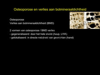
4. Osteoporose en verlies aan botmineraaldichtheid
Osteoporose
el
t uk s
Verlies aan botmineraaldichtheid (BMD)
h
ig r-Y
r
2 vormen van osteoporose / BMD verlies
y le
- gegeneraliseerd: door het hele skelet (heup, LWK)
p u
o G
- gelokaliseerd: in directe nabijheid van gewrichten (hand)
Cheup en onderrug (LWK)
(DEXA) in de
le k
M e
D r.
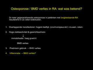
5. Osteoporose / BMD verlies in RA: wat was bekend?
l
2x meer gegeneraliseerde osteoporose in pati谷nten met langbestaande RA,
e
resulterend in 2x vaker botbreuken.
t uk s
h
ig r-Y
1. Overlappende risicofactoren: hogere leeftijd, (postmenopauzale) vrouwen, roken.
r
y le
p u
2. Hoge ziekteactiviteit & gewrichtsschade
o G
C
immobilisatie / laag gewicht
BMD verlies
le k
3.
Me
Prednison gebruik BMD verlies
r.
4. Inflammatie BMD verlies?
D
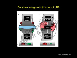
6. Ontstaan van gewrichtsschade in RA
el
t uk s
h
ig r-Y
r
y le
p u
o G
C
le k
Me
D r.
Leidt zelfde mechanisme tot BMD verlies en osteoporose?
Diarra et al, Nat Med 2007
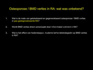
7. Osteoporose / BMD verlies in RA: wat was onbekend?
1.
el
Wat is de mate van gelokaliseerd en gegeneraliseerd osteoporose / BMD verlies
s
in pas gediagnosticeerde RA?
h t uk
ig r-Y
2. Wordt BMD verlies direct veroorzaakt door inflammatoir activiteit in RA?
r
y le
p u
3. Wat is het effect van hedendaagse, moderne behandelstrategie谷n op BMD verlies
o G
in RA?
C
le k
Me
D r.
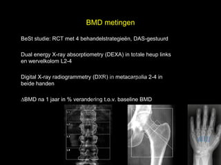
8. BMD metingen
l
BeSt studie: RCT met 4 behandelstrategie谷n, DAS-gestuurd
t uk
Dual energy X-ray absorptiometry (DEXA) in totale heup links
s e
en wervelkolom L2-4
h
ig r-Y
r
y le
p u
Digital X-ray radiogrammetry (DXR) in metacarpalia 2-4 in
o G
beide handen
C
k
BMD na 1 jaar in % verandering t.o.v. baseline BMD
le
Me
D r.
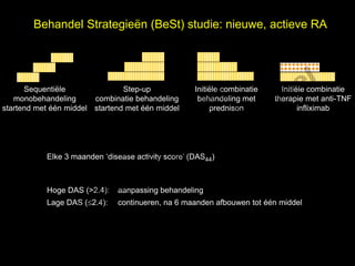
9. Behandel Strategie谷n (BeSt) studie: nieuwe, actieve RA
el
t uk s
h
Sequenti谷le Step-up Initi谷le combinatie Initi谷le combinatie
ig r-Y
monobehandeling combinatie behandeling behandeling met therapie met anti-TNF
r
startend met 辿辿n middel startend met 辿辿n middel prednison infliximab
y le
p u
C o G
k
Elke 3 maanden disease activity score (DAS44)
e le
M
Hoge DAS (>2.4): aanpassing behandeling
r.
Lage DAS (2.4): continueren, na 6 maanden afbouwen tot 辿辿n middel
D
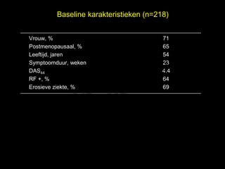
10. Baseline karakteristieken (n=218)
Vrouw, % 71
Postmenopausaal, % 65
el
Leeftijd, jaren
t uk
54
s
Symptoomduur, weken
h
ig r-Y
23
r
DAS44 4.4
RF +, %
y le
p u
64
C o G
Erosieve ziekte, % 69
le k
Me
D r.
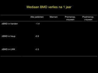
11. Mediaan BMD verlies na 1 jaar
l
Alle pati谷nten Mannen Premenop. Postmenop.
e
vrouwen vrouwen
BMD in handen -1.4
t uk
-1.3 -0.1
s -2.3
h
ig r-Y
r
y le
BMD in heup
p u
o G
-0.9 -1.0 0.1 -1.4
C
le k
BMD in LWK
Me -0.5 -0.1 -0.6 -0.6
D r.
12. Mediaan BMD verlies na 1 jaar
l
Alle pati谷nten Mannen Premenop. Postmenop.
e
vrouwen vrouwen
BMD in handen -1.4
t uk
-1.3 -0.1
s -2.3
h
ig r-Y
r
y le
BMD in heup
p u
o G
-0.9 -1.0 0.1 -1.4
C
le k
BMD in LWK
Me -0.5 -0.1 -0.6 -0.6
D r.
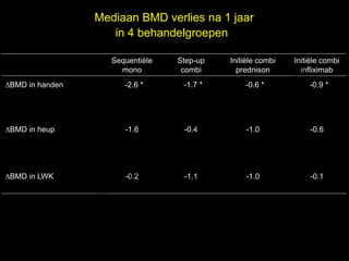
13. Mediaan BMD verlies na 1 jaar
in 4 behandelgroepen
l
Sequenti谷le Step-up Initi谷le combi Initi谷le combi
e
mono combi prednison infliximab
BMD in handen -2.6 *
t uk
-1.7 * -0.6 *
s -0.9 *
h
ig r-Y
r
y le
BMD in heup
p u
o G
-1.6 -0.4 -1.0 -0.6
C
le k
BMD in LWK
Me -0.2 -1.1 -1.0 -0.1
D r.
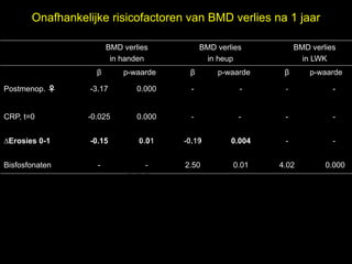
14. Onafhankelijke risicofactoren van BMD verlies na 1 jaar
BMD verlies BMD verlies BMD verlies
in handen in heup in LWK
硫 p-waarde 硫 p-waarde 硫
el p-waarde
t uk s
h
Postmenop. -3.17 0.000 - - - -
r ig r-Y
y le
CRP, t=0 -0.025 0.000 - - - -
p u
o G
C
Erosies 0-1 -0.15 0.01 -0.19 0.004 - -
Bisfosfonaten -
le k - 2.50 0.01 4.02 0.000
Me
D r.
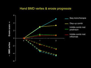
15. Hand BMD verlies & erosie progressie
6
el
Seq.monotherapie
t uk s
Erosie score
h
4 Step-up combi
r ig r-Y
Initi谷le combi met
y le
2 prednison
p u
o G
Initi谷le combi met
C
infliximab
0
k
0 1 2
le
BMD verlies
e
-2
M
-4
D r.
-6
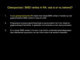
16. Osteoporose / BMD verlies in RA: wat is er nu bekend?
1.
el
In pas gediagnosticeerde RA treedt meer lokaal BMD verlies in handen op dan
s
gegeneraliseerd BMD verlies in heup en LWK.
h t uk
ig r-Y
2. Progressieve erosieve gewrichtsschade is geassocieerd met meer lokaal en
r
gegeneraliseerd botverlies gedeelde mechanismes tussen deze processen.
y le
p u
o G
3. Er is minder BMD verlies in handen in de initi谷le combinatie behandelgroepen
C
door effectievere suppressie van de inflammatie in de eerste 6 maanden.
le k
Me
D r.
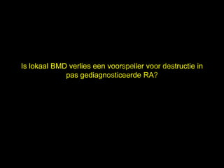
17. el
t uk s
h
ig r-Y
Is lokaal BMD verlies een voorspeller voor destructie in
r
y le
pas gediagnosticeerde RA?
p u
o G
C
le k
Me
D r.
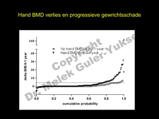
18. Hand BMD verlies en progressieve gewrichtsschade
el
t uk s
h
160
r ig r-Y
No hand BMD loss 0-1 year
y le
Hand BMD loss 0-1 year
40
delta SHS 0-1 year
p u
o G
C
30
k
20
10
e le
M
r.
0
D 0.0 0.2 0.4 0.6
cum ulative probability
0.8 1.0
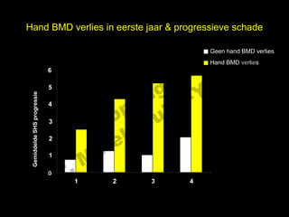
19. Hand BMD verlies in eerste jaar & progressieve schade
l
Geen hand BMD verlies
s e
Hand BMD verlies
t uk
6
h
ig r-Y
r
5
y le
Gemiddelde SHS progressie
p u
4
3
C o G
2
le k
Me
r.
1
D
0
1 2 3 4
follow-up, jaren
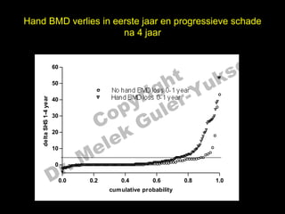
20. Hand BMD verlies in eerste jaar en progressieve schade
na 4 jaar
el
t uk s
60
h
160
ig r-Y
50
r
No hand BMD loss 0-1 year
y le
Hand BMD loss 0-1 year
p u
1-4 year
40
delta SHS 0-1 year
40
30
30
C o G
k
20
le
20
delta
e
10
M
10
r.
0
D
0
0.0
0.0 0.2
0.2 0.4
0.4 0.6
0.6 0.8
0.8 1.0
1.0
cum ulative probability
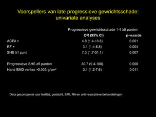
21. Voorspellers van late progressieve gewrichtsschade:
univariate analyses
l
Progressieve gewrichtsschade 1-4 5 punten
t uk
OR (95% CI)
s e
p-waarde
h
ACPA + 4.8 (1.4-16.6) 0.001
r ig r-Y
RF + 3.1 (1.4-6.8) 0.004
y le
SHS 1 punt 7.3 (1.7-31.1) 0.007
p u
o G
C
Progressieve SHS 5 punten 30.7 (9.4-100) 0.000
k
Hand BMD verlies >0.003 g/cm2 3.1 (1.3-7.6) 0.011
e le
M
D r.
Data gecorrigeerd voor leeftijd, geslacht, BMI, RA en anti-resorptieve behandelingen
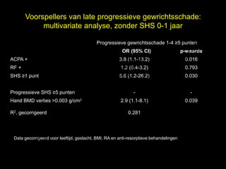
22. Voorspellers van late progressieve gewrichtsschade:
multivariate analyse, zonder SHS 0-1 jaar
l
Progressieve gewrichtsschade 1-4 5 punten
t uk
OR (95% CI)
s e
p-waarde
h
ACPA + 3.8 (1.1-13.2) 0.016
r ig r-Y
RF + 1.2 (0.4-3.2) 0.793
y le
SHS 1 punt 5.6 (1.2-26.2) 0.030
p u
o G
C
Progressieve SHS 5 punten - -
k
Hand BMD verlies >0.003 g/cm2 2.9 (1.1-8.1) 0.039
R2, gecorrigeerd
e le 0.281
M
D r.
Data gecorrigeerd voor leeftijd, geslacht, BMI, RA en anti-resorptieve behandelingen
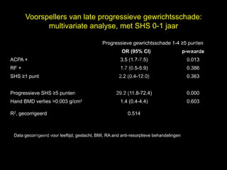
23. Voorspellers van late progressieve gewrichtsschade:
multivariate analyse, met SHS 0-1 jaar
l
Progressieve gewrichtsschade 1-4 5 punten
t uk
OR (95% CI)
s e
p-waarde
h
ACPA + 3.5 (1.7-7.5) 0.013
r ig r-Y
RF + 1.7 (0.5-5.9) 0.386
y le
SHS 1 punt 2.2 (0.4-12.0) 0.363
p u
o G
C
Progressieve SHS 5 punten 29.2 (11.8-72.4) 0.000
k
Hand BMD verlies >0.003 g/cm2 1.4 (0.4-4.4) 0.603
R2, gecorrigeerd
e le 0.514
M
D r.
Data gecorrigeerd voor leeftijd, geslacht, BMI, RA and anti-resorptieve behandelingen
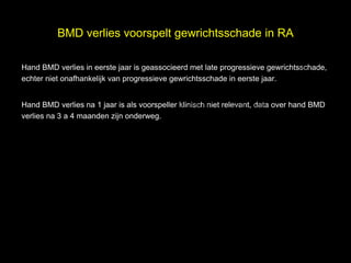
24. BMD verlies voorspelt gewrichtsschade in RA
l
Hand BMD verlies in eerste jaar is geassocieerd met late progressieve gewrichtsschade,
echter niet onafhankelijk van progressieve gewrichtsschade in eerste jaar.
t uk s e
h
ig r-Y
r
Hand BMD verlies na 1 jaar is als voorspeller klinisch niet relevant, data over hand BMD
y le
verlies na 3 a 4 maanden zijn onderweg.
p u
o G
C
le k
Me
D r.
25. el
t uks
BMD verliesgh
ri r-Y
in OA
p uy le
C o G
le k
Me
D r.
26. Pathogenese OA
el
Multifactorieel:
s
Degeneratieve factoren
Biomechanische factoren
h t uk
ig r-Y
Metabole factoren
Hormonale factoren
r
y le
p u
Genetische factoren
o G
Inflammatoire factoren?
C
le k
Me
D r.
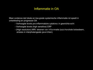
27. Inflammatie in OA
el
Meer evidence dat lokale en low-grade systemische inflammatie rol speelt in
s
ontwikkeling en progressie OA
h t uk
- Verhoogde levels pro-inflammatoire cytokines in gewrichtsvocht
ig r-Y
- Verhoogde levels (high sensitive) CRP
r
- (High resolution) MRI: tekenen van inflammatie (subchondrale botoedeem,
y le
p u
erosies in interphalangeale gewrichten)
C o G
le k
Me
D r.
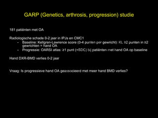
28. GARP (Genetics, arthrosis, progression) studie
l
181 pati谷nten met OA
t uk
Radiologische schade 0-2 jaar in IPJs en CMC1
s e
h
- Baseline: Kellgren-Lawrence score (0-4 punten per gewricht): KL 2 punten in 2
ig r-Y
gewrichten = hand OA
r
- Progressie: OARSI atlas: 1 punt (=SDC) bij pati谷nten met hand OA op baseline
y le
p u
o G
Hand DXR-BMD verlies 0-2 jaar
C
le k
Vraag: Is progressieve hand OA geassocieerd met meer hand BMD verlies?
Me
D r.
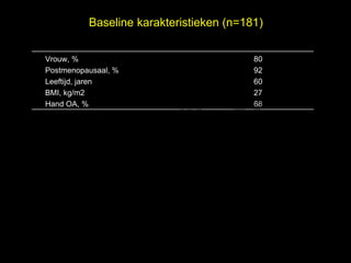
29. Baseline karakteristieken (n=181)
Vrouw, % 80
Postmenopausaal, % 92
el
t uk s
Leeftijd, jaren 60
h
BMI, kg/m2 27
ig r-Y
Hand OA, % 68
r
y le
p u
o G
C
le k
Me
D r.
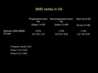
30. BMD verlies in OA
el
Progressieve hand Non-progressieve hand Geen hand OA
s
OA OA
h
Groep 1 n=39
t uk Groep 2 n=84 Groep 3 n=58
Mediaan (IQR) BMD
r ig r-Y
y le
-2.2% -1.4% -1.4%
p u
0-2 jaar (-4.1 tot -1.4) (-2.9 tot -0.6) (-3.1 tot -0.4)
C o G
P-waarde, overall: 0.032
le k
Groep 1 vs 2: 0.045
Me
r.
Groep 2 vs 3: 0.804
D
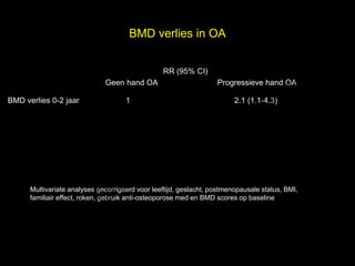
31. BMD verlies in OA
RR (95% CI)
el
t uk s
Geen hand OA Progressieve hand OA
BMD verlies 0-2 jaar 1
h
ig r-Y
2.1 (1.1-4.3)
r
y le
p u
o G
C
le k
Me
Multivariate analyses gecorrigeerd voor leeftijd, geslacht, postmenopausale status, BMI,
r.
familiair effect, roken, gebruik anti-osteoporose med en BMD scores op baseline
D
32. BMD verlies in OA
l
Lokaal BMD verlies in handen is geassocieerd met (progressieve) hand OA na 2
e
t uk s
jaar.
h
ig r-Y
r
Pathogenese van OA is multifactorieel, mogelijk speelt (lokaal) inflammatoir
activiteit ook een rol.
y le
p u
C o G
le k
Me
D r.
33. el
t uks
IWO, dank voorgh prijs
ri r-Y
HvP
p uy le
C o G
le k
M e
D r.