


More Related Content
What's hot (20)
Viewers also liked (20)


Similar to Vurdering av og for l├”ring (20)


More from Eva Bratvold (20)


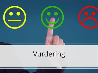
Vurdering av og for l├”ring
- 1. Vurdering
- 2. Hvorfor vurdering? ŌĆó Vurdering skal minske gap mellom n├źsituasjon og ├Ėnsket m├źloppn├źelse. ŌĆó For at alle skal vokse opp kreves det at vi gj├Ėr hullene mindre og at de f├źr nok mat slik at de ikke faller gjennom senere ŌĆó P├ź den m├źten skal vi sikre at alle kommer trygt fram og ikke faller gjennom underveis ŌĆó Hva gj├Ėr du hvis dine elever ikke har vokst seg store nok ŌĆō og risikerer ├ź falle gjennom hullene?
- 3. ŌĆóMange elever f├Ėler at skolen best├źr av ├ź gj├Ėre oppgaver og aktiviteter ŌĆó uten at de vet hvorfor ŌĆō de forst├źr ikke relevansen ŌĆó som det er vanskelig ├ź vite om de lykkes med eller ikke ŌĆó fordi de ikke vet hva som er viktig, og hvordan de blir vurdert
- 4. Formativ og summativ vurdering Hva er forskjellene?
- 5. Summativ vurdering ŌĆó Vurdering av l├”ring (VAL) ŌĆó Omtrent som en obduksjon ŌĆō vi kan finne ut hva pasienten d├Ėde av, men det er for sent ├ź gj├Ėre noe med det ŌĆó P├ź samme m├źte som en lege b├Ėr vurdere om vurderingen av hva som feilte pasienten var korrekt, og at riktig behandling ble gitt - for ├ź l├”re av sine feil og l├”re av situasjonen - b├Ėr l├”reren se p├ź elevenes resultater som en evaluering av sin egen undervisningspraksis og evt. endre kurs
- 6. Formativ vurdering ŌĆó Vurdering for l├”ring (VFL) ŌĆó Omtrent som en helsesjekk hos legen ŌĆō hvor man f├źr avdekket hvordan helsesituasjonen er, og f├źr behandling, medisin eller r├źd for ├ź holde seg sunn og frisk slik at man kan utvikle seg videre med god livskvalitet ŌĆó Som legen m├ź unders├Ėke pasienten for ├ź foreskrive riktig kur, m├ź l├”reren vite mange ting om sin elev ŌĆō slik at man vet hva eleven trenger for ├ź komme videre
- 7. Trener eller dommer? ŌĆó N├źr man arbeider med summativ vurdering er man som en dommer, mens med formativ vurderinger er man mer som en trener ŌĆó Dessverre kan elever oppfatte en som en dommer n├źr man ├Ėnsker ├ź v├”re en trener ŌĆō og de vil pr├Ėve ├ź skjule det de ikke kan framfor ├ź vise hva de trenger hjelp til for ├ź bli bedre ŌĆó Det er derfor viktig at man har en forst├źelse og en atmosf├”re som gj├Ėr at man forst├źr at tilbakemeldingene gj├Ėres som en trener ŌĆō ikke som en dommer
- 8. Vurdering for l├”ring dreier seg om ŌĆó L├”replanforst├źelse ŌĆó Naturlig progresjon ŌĆó Forst├źelige l├”ringsm├źl ŌĆó Gjenkjenne at m├źl er n├źdd ŌĆó Forholdet mellom oppgaver og kompetanse/l├”ringsm├źl ŌĆó Elevene m├ź forst├ź vurderingskriteriene ŌĆó Fokus p├ź l├”ring ŌĆó Relasjoner og interaksjoner i klasserommet (ogs├ź peer-to-peer) ŌĆó ├ģ forst├ź hvor de l├”rende befinner seg (hva som mestres, ikke mestres) ŌĆó ├ģ gi hjelp for at de skal komme videre
- 9. Begreper ŌĆó Feedback, tilbakemeldinger, kan skje p├ź flere m├źter ŌĆó Som formativ vurdering i form av en underveisvurdering, som kan v├”re en fremovermelding (feed forward) eller peke mot overordnet m├źl (feed up) ŌĆó Som summativ vurdering, f.eks. i form av en karakter eller poenggivning Kanskje vi kan si at vurdering handler om ├ź gj├Ėre umulig om til mulig?
- 11. Elever l├”rer best n├źr de ŌĆó Forst├źr hva de skal l├”re, og hva som blir forventet (l├”ringsm├źl) ŌĆó F├źr tilbakemeldinger som forteller om kvaliteten p├ź arbeidet (kjennetegn p├ź m├źloppn├źelse) ŌĆó F├źr r├źd om hvordan de kan gj├Ėre det bedre (suksesskriterier) ŌĆó Er involverte i eget l├”ringsarbeid, f.eks. vurdere eget arbeid og utvikling (egenvurdering)
- 12. God vurdering ŌĆó Effektive fremovermeldinger ŌĆó Konstruktive ŌĆó Aktivt involverte elever ŌĆó Dekonstruksjon ŌĆó Utvikle kriterier ŌĆó Elevene vurderer seg selv og forst├źr hvordan de skal forberede seg ŌĆó Vurderingskriterier ŌĆó L├”re ├ź l├”re, l├”ringsstrategier ŌĆó Lese med hensikt ŌĆó Timing ŌĆó Undervisning tilpasset resultat fra vurderingen ŌĆó Bevissthet om vurderingens rolle for motivasjon og selvf├Ėlelse ŌĆó Klare m├źl ŌĆó Umiddelbar respons ŌĆó Utfordringer tilpasset niv├ź ŌĆó F├Ėlelse av kontroll og mestring ŌĆó Individuell eller sosial dimensjon
- 13. Fremovermeldinger ŌĆó Fremovermeldinger (feed forward) m├ź v├”re konstruktiv kritikk ŌĆō at elevene f├źr hjelp til ├ź forbedre og komme videre ŌĆó Fokus p├ź forbedringer fra tidligere (progresjon, evne til ├ź ta til seg tidligere tilbakemeldinger og endre produkt) ŌĆó Fokus p├ź kvaliteter i produktet (faglig ros) ŌĆó Fokus p├ź veien videre (konstruktiv kritikk, strategier)
- 14. Vurderingskriterier ŌĆó Kriteriene m├ź v├”re objektive, ikke subjektive. L├”rer oppfattes som subjektiv dersom vurderingskriterier mangler (eleven oppfatter seg som problem, ikke oppgaven) ŌĆó M├ź tilbakemeldingene alltid v├”re positive, eller kan dette f├Ėre til at man gir mest ros og lite nyttige (konstruktive) tilbakemeldinger? ŌĆó Det er en forskjell p├ź tilbakemelding basert p├ź mestringsniv├ź og tilbakemelding basert p├ź engasjement i l├”ringsaktiviteten ŌĆó I bruk av medelevvurdering (peer-to- peer) er faren at tilbakemeldingene blir for snille eller at de er u├”rlige. Noen kan ┬½straffe┬╗ medelever gjennom negative tilbakemeldinger.
- 15. M├źl og kriterier ŌĆó Noen ganger m├ź man zoome ut for ├ź f├ź oversikt. Det er ikke alltid s├ź enkelt ├ź forst├ź hvorfor man skal holde p├ź med ulike oppgaver om man ikke ser hvor det f├Ėrer en ŌĆō og det har betydning b├źde for motivasjon og relevans. ŌĆó Tenk omtrent som om du st├źr inne i en hekklabyrint. Da ser du bare de n├”rmeste hekkene, men du aner ikke hvordan du skal komme fram til m├źlet. Det er lettere hvis du ser hele labyrinten ovenfra ŌĆō og da kan du planlegge ruta uten ├ź g├ź feil for mange ganger.
- 16. Vurderinger ŌĆó Det er ikke kvantiteten, men kvaliteten p├ź dine tilbakemeldinger som har betydning ŌĆó Tenk over; hjelper din tilbakemelding eleven med ├ź l├”re? ŌĆó Hva skal de bruke tiden til og hva f├Ėrer det til? ŌĆó Det som har betydning er informasjon om kvalitet p├ź elevens arbeid, prosesser/strategier og hva som m├ź til for ├ź forbedre resultatet og komme videre ŌĆó Rangering i form av karakterer og poeng hjelper lite ŌĆō og v├”r oppmerksom p├ź om du egentlig utf├Ėrer sm├ź summative vurderinger framfor formative ŌĆó Personrettet ros, ris og r├źd fungerer d├źrlig og er ikke l├”ringsfremmende. Det kan ogs├ź virke mot sin hensikt. Man m├ź heller anerkjenne innsats og faglig kvalitet, ikke fokusere p├ź person
- 17. D├źrlige tilbakemeldinger ŌĆó Det er mange tilbakemeldinger som ikke hjelper; ŌĆó Blir fortalt at man kan gj├Ėre noe bedre, selv om man selv f├Ėler at man har gjort sitt beste ŌĆó Blir fortalt ├ź jobbe mer og bedre ŌĆō men ikke hvordan ŌĆó F├źr tilbakemeldinger p├ź slutten eller etter en arbeidsprosess, kan ikke bruke informasjonen (timing ŌĆō tilbakemelding kommer for sent) ŌĆó F├źr personrettet ros, men ikke hjelp til ├ź komme videre faglig (bra jobba!) ŌĆó F├źr beskjed om at oppgaven er best├źtt, eller at man har f├źtt en karakter (summativ vurdering) ŌĆō men ingen ├Ėvrig informasjon
- 18. Gode tilbakemeldinger ŌĆó Mark Barnes mener at dette er kjennetegn p├ź gode tilbakemeldinger (summarize, explain, redirict, resubmit): ŌĆó Fortelle spesifikt hva eleven har gjort (oppsummering) ŌĆó Forklare hva man har forst├źtt og ikke (forst├źelse, avklar misforst├źelser) ŌĆó Dirigere i riktig retning, forklare og hvordan det har betydning for hva som skal gj├Ėres. Bygg p├ź elevens forutsetninger. ŌĆó Be om ├ź f├ź se det nye arbeidet/produktet n├źr det er endret (viser at man er interessert og gir forventning om at eleven begynner ├ź arbeide igjen)
- 19. Tilbakemeldinger ŌĆó Elevene m├ź se at det er en sammenheng mellom innsats og faglig fremgang ŌĆó Elevene m├ź tro det er mulig ├ź komme videre ŌĆó Rangering og karakterer f├Ėrer ofte til at de sammenligner seg med andre, og det f├Ėrer til at de viktige tingene (hvilken framgang har du hatt, progresjon, og hvordan du kan komme videre) ikke blir brukt/kommer fram ŌĆó ├ģ f├ź en karakter kan prege framtidig utf├Ėrelse, man har blitt satt i en b├źs ŌĆō noe som f├Ėrer til at det er vanskelig ├ź f├ź l├Ėftet eleven opp p├ź et h├Ėyere niv├ź ŌĆó Elevene kan tenke tilbakemeldingen som ren kritikk, mens l├”reren har tenkt den som konstruktiv kritikk ŌĆō elevene ser hva tilbakemelding er p├ź en bestemt oppgave, l├”rer tenker i et lengre tidsspenn ŌĆó ├ģ rette store bunker og gi summative vurderinger hjelper lite, bruk heller tid p├ź underveisvurderinger ŌĆó Det er ikke vurderingen som skal v├”re bra (den skal hjelpe) ŌĆō husk ├ź v├”re trener, ikke dommer
- 20. Ulike niv├źer ŌĆó Oppgaveniv├ź (oppgave, produkt, feil, mangler, utseende) ŌĆó Prosessniv├ź (fokuserer p├ź prosesser og l├”ringsstrategier for ├ź forbedre oppgave, produkt og l├”ringsprosess) ŌĆó Selvregulering (bevisstgj├Ėring av eleven ŌĆō hva gj├Ėr eleven n├źr de skal i gang med et arbeid, reflektere over egen kompetanse og hvordan man handler; hvor er jeg, hvor skal jeg, hvordan kommer jeg dit) metaforst├źelse p├ź prosesser, kjenne til m├źlet (hva skal jeg l├”re), suksesskriterier (kjennetegn p├ź m├źloppn├źelse) ŌĆō f├Ėrst da f├źr man retningen og kan finne ut i hvilken grad man har lyktes ŌĆó Personniv├ź ŌĆō informasjon blir rettet mot person, tar fokus vekk fra arbeidsprosess/oppgave, dyrker fram unnvikelsesstrategier (vi ├Ėnsker jo mestring, unng├źr oppgaven hvis vi ikke tror vi kan mestre den). Dette niv├źet b├Ėr unng├źs. Oftest gis tilbakemeldinger p├ź oppgaveniv├ź ŌĆō det gj├Ėr det vanskelig ├ź overf├Ėre informasjon til andre kontekster. Hvorfor er det oftest dette man gj├Ėr? Kilde: Hattie og Timperley
- 21. Aktivt engasjerte elever ŌĆó Hvordan vet man at man kan det man skal? ŌĆó La elevene v├”re med p├ź ├ź utforme kriterier. Her er to metoder; ŌĆó Dekonstruksjon ŌĆō dette er et eksempel p├ź en god besvarelse/produkt ŌĆō hvorfor er besvarelsen/produktet bra? Bruk kriteriene man finner til ├ź lage ny besvarelse eller nytt produkt ŌĆó Placemat ŌĆō elever i grupper skriver hver for seg hva de mener kan v├”re gode kriterier. Deretter diskuterer man i gruppa hvilke av kriteriene man kan v├”re enige om
- 22. Involverte elever ŌĆó De m├ź vite hvor de er ŌĆō hva de kan, hva de har l├”rt ŌĆó De m├ź vite hvor de skal ŌĆō hva de skal kunne, hva de skal l├”re ŌĆó De m├ź kunne sammenligne det de kan n├ź og m├źlet for ├ź finne gapet ŌĆó De m├ź kunne tette gapet ŌĆō i samarbeid med l├”rer
- 23. Endret undervisning ŌĆó L├”reren m├ź evaluere sin undervisning basert p├ź elevenes resultater, og eventuelt endre kurs ŌĆó Det er derfor viktig ├ź overv├źke progresjon og forst├źelse ŌĆó Forst├źr elevene hva de skal gj├Ėre (oppgave, relevans) ŌĆó Forst├źr elevene hvordan de skal jobbe (strategi, suksesskriterier) ŌĆó Klarer de ├ź l├Ėse oppgavene eller forst├ź konseptene (underveisvurdering) ŌĆó En metode for ├ź planlegge med innlagte sjekkpunkt er baklengs planlegging (backward design) ŌĆō slik at man f├źr endret undervisningen i tide
- 24. Timing ŌĆó N├źr skal man vurdere? Vurdere f├Ėr det er for sent! Man m├ź gi underveisvurdering n├źr det er nyttig ŌĆō i prosessen ŌĆō ikke n├źr man er ferdig. ŌĆó Rett etter framf├Ėring gj├Ėr at man ikke f├źr endret noe ŌĆō bedre ├ź v├”re tilstede p├ź generalpr├Ėve ŌĆó Enkelttimer, perioder, hele fagl├Ėpet ŌĆō tidsperioden man tenker har betydning for n├źr og hva man skal vurdere ŌĆó Raske tilbakemeldinger er mest effektivt ŌĆó P├ź hvilke m├źter kan du s├Ėrge for at elevene f├źr raske tilbakemeldinger mens de er i arbeidsprosessen?
- 25. Forst├źr elevene hva det betyr og hvorfor de skal l├”re det? Tydelige l├”ringsm├źl og vurderingskriteri er, Endre og/eller forklare bedre Elevene kan svare p├ź; Hva gj├Ėr du n├ź? Hvorfor gj├Ėr du dette? Hvordan henger dette sammen med det du skal kunne n├źr du er ferdig med utdanningen? Undervisning p├źg├źr, elevene jobber med oppgaver og aktiviteter Vurdering for l├”ring Star t Underveis - vurdering, framover- meldinger NEI JA Kan elevene ta del I arbeidet med ├ź utforme Fokus p├ź prosess og strategier Fokus p├ź oppgave/arb eid Det spesifikke i oppgaven (feiltolking ) Kvalitet p├ź arbeidet For ├ź l├Ėse og forbedre oppgave og behandling av informasjo n Strategi- bruk/ l├”rings- prosesser Fokus p├ź egenregulerin g Hvor er jeg og hvordan kommer jeg videre Timing