3.acquired valvular heart disease
5 likes483 views

The document describes x-ray findings of mitral stenosis of varying severity. For mild mitral stenosis, chest x-rays may be normal but show an enlarged left atrium. Moderate to severe mitral stenosis is seen as an enlarged left atrium, elevated left main bronchus, and displaced descending aorta on chest x-rays. Severe mitral stenosis additionally shows enlarged pulmonary arteries and veins as well as displacement of the esophagus. Differential diagnoses include pectus excavatum and partial absence of the pericardium.


More Related Content
Viewers also liked (20)
More from Ngoan Pham (8)


3.acquired valvular heart disease
- 1. 1 CHA├ģN ├æOA├ÖN H├īNH A├øNH BE├äNH VAN TIM MA├ēC PHA├øI BS.NGUYE├āN QUY├Ö KHOA├ÖNG BS.NGUYE├āN QUANG TRO├ÅNG 6/19/2013
- 2. 2 DA├śN BA├śI He├»p van 2 la├╣. ├æa├»i c├Č├┤ng. X quang He├»p van 2 la├╣ nhe├». X quang He├»p van 2 la├╣ trung b├¼nh-na├½ng. Thay ├▒o├źi ve├Ā tim. Thay ├▒o├źi ve├Ā ma├»ch ma├╣u. Thay ├▒o├źi ve├Ā chu├╗ mo├ó pho├źi. 6/19/2013
- 3. 3 DA├śN BA├śI H├┤├╗ van 2 la├╣. ├æa├»i c├Č├┤ng. X quang H├┤├╗ van 2 la├╣ nhe├». X quang H├┤├╗ van 2 la├╣ trung b├¼nh. X quang He├»p van 2 la├╣ na├½ng. Ca├╣c the├ź ke├Īt h├┤├»p. 6/19/2013
- 4. 4 DA├śN BA├śI He├»p van ├æo├żng ma├»ch chu├╗. ├æa├»i c├Č├┤ng. X quang. Co├żng h├Č├┤├╗ng t├Č├Ė. 6/19/2013
- 5. 5 DA├śN BA├śI H├┤├╗ van ├æo├żng ma├»ch chu├╗. ├æa├»i c├Č├┤ng. X quang. Chu├»p co├╣ ca├╗n quang. Co├żng h├Č├┤├╗ng t├Č├Ė. 6/19/2013
- 6. 6 DA├śN BA├śI Be├żnh ly├╣ van ├æo├żng ma├»ch pho├źi. Be├żnh ly├╣ van 3 la├╣. ├¢├Öng du├»ng la├óm sa├Ėng. Ke├Īt lua├żn. 6/19/2013
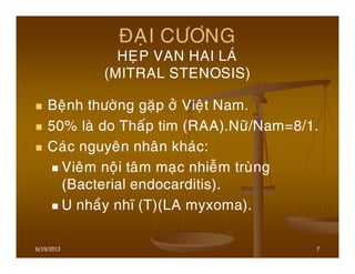
- 7. 7 ├æA├ÅI C├¢├öNG HE├ÅP VAN HAI LA├Ö (MITRAL STENOSIS) Be├żnh th├Č├┤├Ėng ga├½p ├┤├╗ Vie├żt Nam. 50% la├Ė do Tha├Īp tim (RAA).N├Č├Ą/Nam=8/1. Ca├╣c nguye├ón nha├ón kha├╣c: Vie├óm no├żi ta├óm ma├»c nhie├Żm tru├Ėng (Bacterial endocarditis). U nha├Āy nh├│ (T)(LA myxoma). 6/19/2013
- 8. 8 ÑAÏI CÛÔNG HEÏP VAN HAI LAÙ 6/19/2013
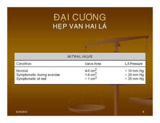
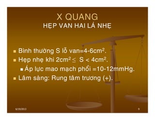
- 9. 9 X QUANG HE├ÅP VAN HAI LA├Ö NHE├Å B├¼nh th├Č├┤├Ėng S lo├Ż van=4-6cm2. He├»p nhe├» khi 2cm2 Ōēż S < 4cm2. A├Öp l├Č├»c mao ma├»ch pho├źi =10-12mmHg. La├óm sa├Ėng: Rung ta├óm tr├Č├┤ng (+). 6/19/2013
- 10. 10 X QUANG HEÏP VAN HAI LAÙ NHEÏ NORMAL MITRAL VALVE MITRAL STENOSIS 6/19/2013
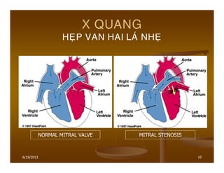
- 11. 11 X QUANG HE├ÅP VAN HAI LA├Ö NHE├Å X quang: Bo├╣ng tim ch├Ča tha├Īy thay ├▒o├źi g├¼. Pha├ón bo├Ī ma├»ch ma├╣u pho├źi cu├Ąng ch├Ča tha├Īy thay ├▒o├źi. Do va├ży,X quang tim-pho├źi b├¼nh th├Č├┤├Ėng kho├óng loa├»i tr├Č├Ė ├▒├Č├┤├»c He├»p van hai la├╣. 6/19/2013
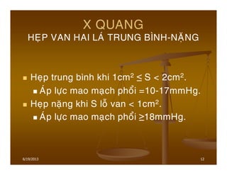
- 12. 12 X QUANG HE├ÅP VAN HAI LA├Ö TRUNG B├īNH-NA├ŗNG He├»p trung b├¼nh khi 1cm2 Ōēż S < 2cm2. A├Öp l├Č├»c mao ma├»ch pho├źi =10-17mmHg. He├»p na├½ng khi S lo├Ż van < 1cm2. A├Öp l├Č├»c mao ma├»ch pho├źi Ōēź18mmHg. 6/19/2013
- 13. 13 X QUANG HE├ÅP VAN HAI LA├Ö TRUNG B├īNH-NA├ŗNG Sinh ly├╣ be├żnh: Ta├®c nghe├Ąn do├Ėng cha├╗y t├Č├Ė Nh├│ (T) xuo├Īng Tha├Īt (T) Ta├¬ng a├╣p l├Č├»c Nh├│ (T) Da├Ąn buo├Āng Nh├│ (T) Ta├¬ng a├╣p l├Č├»c TM pho├źi Ta├¬ng a├╣p l├Č├»c ├æM pho├źi Ta├¬ng a├╣p l├Č├»c Tha├Īt (P) Gia├Ąn buo├Āng Tha├Īt (P). 6/19/2013
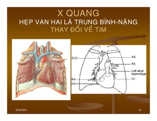
- 14. 14 X QUANG HE├ÅP VAN HAI LA├Ö TRUNG B├īNH-NA├ŗNG THAY ├æO├ģI VE├Ć TIM Da├Ąn buo├Āng Nh├│ (T) khi ├▒├Č├┤├Ėng k├Łnh Nh├│ (T)>7cm (├▒o t├Č├Ė b├┤├Ė d├Č├┤├╣i PQ go├Īc (T) ├▒e├Īn b├┤├Ė pha├╗i Nh├│ (T)). Phe├Ī qua├╗n go├Īc (T) b├▓ ├▒a├źy le├ón cao. Co├╣ h├¼nh a├╗nh b├┤├Ė ├▒o├ói ├┤├╗ b├┤├Ė (P) cu├╗a tim. 6/19/2013
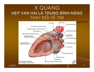
- 15. 15 X QUANG HE├ÅP VAN HAI LA├Ö TRUNG B├īNH-NA├ŗNG THAY ├æO├ģI VE├Ć TIM ├ægMC xuo├Īng b├▓ ├▒a├źy qua (T). Phim tha├║ng co├╣ uo├Īng Baryte:Th├Č├»c qua├╗n co├╣ the├ź b├▓ ├▒a├źy le├żch,th├Č├┤├Ėng la├Ė qua (P),├▒o├ói khi b├▓ ├▒a├źy le├żch qua (T). Phim che├Īch tr├Č├┤├╣c (P)(RAO) co├╣ uo├Īng Baryte:Th├Č├»c qua├╗n b├▓ ├▒a├źy ra sau ro├Ą nha├Īt. 6/19/2013
- 16. 16 X QUANG HE├ÅP VAN HAI LA├Ö TRUNG B├īNH-NA├ŗNG THAY ├æO├ģI VE├Ć TIM 6/19/2013
- 17. 17 X QUANG HE├ÅP VAN HAI LA├Ö TRUNG B├īNH-NA├ŗNG THAY ├æO├ģI VE├Ć TIM 6/19/2013
- 18. 18 X QUANG HE├ÅP VAN HAI LA├Ö TRUNG B├īNH-NA├ŗNG THAY ├æO├ģI VE├Ć TIM 6/19/2013
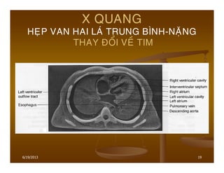
- 19. 19 X QUANG HE├ÅP VAN HAI LA├Ö TRUNG B├īNH-NA├ŗNG THAY ├æO├ģI VE├Ć TIM 6/19/2013
- 20. 20 X QUANG HE├ÅP VAN HAI LA├Ö TRUNG B├īNH-NA├ŗNG THAY ├æO├ģI VE├Ć TIM LEFT ATRIAL ENLARGEMENT is best confirmed by measuring the distance from the midinferior border of the left main bronchus to the right lateral border of the left atrial density. -This distance is less than 7cm in 90% of normal patients and is greater than 7cm in 90% of left atrial enlargement patients,as proven by echocardiography. -This measurement can be approximated by placing oneŌĆÖs right fifth finger under the left bronchus,and while keeping the fingers closed,if the left atrium is seen beyond oneŌĆÖs four fingertips,the left atrium is enlarged. 6/19/2013
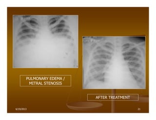
- 21. 21 PULMONARY EDEMA / MITRAL STENOSIS AFTER TREATMENT 6/19/2013
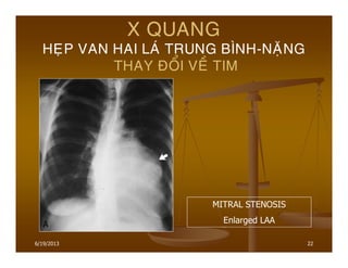
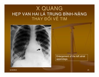
- 22. 22 X QUANG HE├ÅP VAN HAI LA├Ö TRUNG B├īNH-NA├ŗNG THAY ├æO├ģI VE├Ć TIM MITRAL STENOSIS Enlarged LAA 6/19/2013
- 23. 23 X QUANG HE├ÅP VAN HAI LA├Ö TRUNG B├īNH-NA├ŗNG THAY ├æO├ģI VE├Ć TIM Enlargement of the left atrial appendage. 6/19/2013
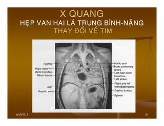
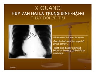
- 24. 24 X QUANG HE├ÅP VAN HAI LA├Ö TRUNG B├īNH-NA├ŗNG THAY ├æO├ģI VE├Ć TIM -Elevation of left main bronchus. -Double shadow of the large left atrium (arrow). -Right atrial border is limited below by the entry of the inferior vena cava. 6/19/2013
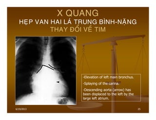
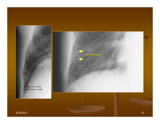
- 25. 25 X QUANG HE├ÅP VAN HAI LA├Ö TRUNG B├īNH-NA├ŗNG THAY ├æO├ģI VE├Ć TIM -Elevation of left main bronchus. -Splaying of the carina. -Descending aorta (arrow) has been displaced to the left by the large left atrium. 6/19/2013
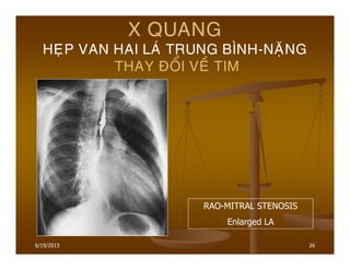
- 26. 26 X QUANG HE├ÅP VAN HAI LA├Ö TRUNG B├īNH-NA├ŗNG THAY ├æO├ģI VE├Ć TIM RAO-MITRAL STENOSIS Enlarged LA 6/19/2013
- 27. 27 X QUANG HE├ÅP VAN HAI LA├Ö TRUNG B├īNH-NA├ŗNG THAY ├æO├ģI VE├Ć TIM Ca├Ėng nga├Ėy Nh├│ (T) ca├Ėng to ra,nha├Īt la├Ė khi co├╣ Rung nh├│.Nh├│ (T)co├╣ the├ź v├Č├┤├»t ra ngoa├Ėi b├┤├Ė Nh├│ (P). Cung ├æMP cu├Ąng to ra do ta├¬ng a├╣p trong buo├Āng Tha├Īt (P). 6/19/2013
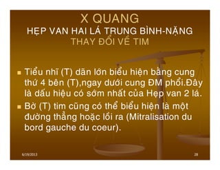
- 28. 28 X QUANG HE├ÅP VAN HAI LA├Ö TRUNG B├īNH-NA├ŗNG THAY ├æO├ģI VE├Ć TIM Tie├źu nh├│ (T) da├Ąn l├┤├╣n bie├źu hie├żn ba├©ng cung th├Č├╣ 4 be├ón (T),ngay d├Č├┤├╣i cung ├æM pho├źi.├æa├óy la├Ė da├Īu hie├żu co├╣ s├┤├╣m nha├Īt cu├╗a He├»p van 2 la├╣. B├┤├Ė (T) tim cu├Ąng co├╣ the├ź bie├źu hie├żn la├Ė mo├żt ├▒├Č├┤├Ėng tha├║ng hoa├½c lo├Āi ra (Mitralisation du bord gauche du coeur). 6/19/2013
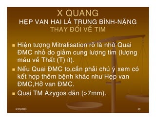
- 29. 29 X QUANG HE├ÅP VAN HAI LA├Ö TRUNG B├īNH-NA├ŗNG THAY ├æO├ģI VE├Ć TIM Hie├żn t├Č├┤├»ng Mitralisation ro├Ą la├Ė nh├┤├Ė Quai ├æMC nho├╗ do gia├╗m cung l├Č├┤├»ng tim (l├Č├┤├»ng ma├╣u ve├Ā Tha├Īt (T) ├Łt). Ne├Īu Quai ├æMC to,ca├Ān pha├╗i chu├╣ y├╣ xem co├╣ ke├Īt h├┤├»p the├óm be├żnh kha├╣c nh├Č He├»p van ├æMC,H├┤├╗ van ├æMC. Quai TM Azygos da├Ąn (>7mm). 6/19/2013
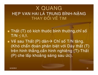
- 30. 30 X QUANG HE├ÅP VAN HAI LA├Ö TRUNG B├īNH-NA├ŗNG THAY ├æO├ģI VE├Ć TIM Tha├Īt (T) co├╣ k├Łch th├Č├┤├╣c b├¼nh th├Č├┤├Ėng,ch├” so├Ī T/N Ōēż 0,5. Ve├Ā sau Tha├Īt (P) da├Ąn Ch├” so├Ī T/N ta├¬ng. (Kho├╣ cha├źn ├▒oa├╣n pha├ón bie├żt v├┤├╣i Da├Ėy tha├Īt (T) tre├ón h├¼nh tha├║ng,ca├Ān h├¼nh nghie├óng (T)-Tha├Īt (P) che la├Īp khoa├╗ng sa├╣ng sau ├Č├╣c). 6/19/2013
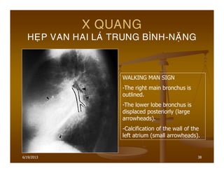
- 31. 6/19/2013 31 X QUANG HE├ÅP VAN HAI LA├Ö TRUNG B├īNH-NA├ŗNG THAY ├æO├ģI VE├Ć TIM H├¼nh a├╗nh ├Łt ga├½p la├Ė vo├ói ho├╣a van hai la├╣(40%), vo├Ėng van 2 la├╣(10%) va├Ė ├┤├╗ tha├Ėnh Nh├│ (T). Phim nghie├óng (T):Nh├│ (T) to ├▒a├źy PQ go├Īc (T) ra sau (Walking man sign).
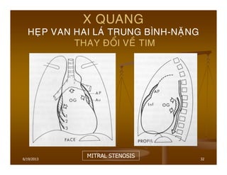
- 32. 6/19/2013 32 X QUANG HE├ÅP VAN HAI LA├Ö TRUNG B├īNH-NA├ŗNG THAY ├æO├ģI VE├Ć TIM MITRAL STENOSIS
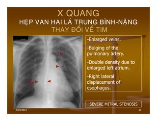
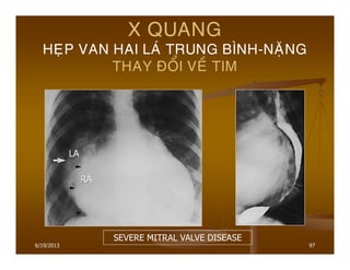
- 33. 6/19/2013 33 X QUANG HE├ÅP VAN HAI LA├Ö TRUNG B├īNH-NA├ŗNG THAY ├æO├ģI VE├Ć TIM -Enlarged veins. -Bulging of the pulmonary artery. -Double density due to enlarged left atrium. -Right lateral displacement of esophagus. SEVERE MITRAL STENOSIS
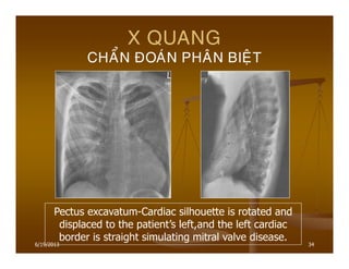
- 34. 6/19/2013 34 X QUANG CHA├ģN ├æOA├ÖN PHA├éN BIE├äT Pectus excavatum-Cardiac silhouette is rotated and displaced to the patientŌĆÖs left,and the left cardiac border is straight simulating mitral valve disease.
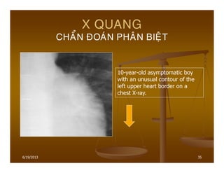
- 35. 6/19/2013 35 X QUANG CHA├ģN ├æOA├ÖN PHA├éN BIE├äT 10-year-old asymptomatic boy with an unusual contour of the left upper heart border on a chest X-ray.
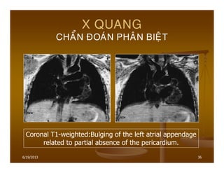
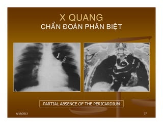
- 36. 6/19/2013 36 X QUANG CHA├ģN ├æOA├ÖN PHA├éN BIE├äT Coronal T1-weighted:Bulging of the left atrial appendage related to partial absence of the pericardium.
- 37. 6/19/2013 37 X QUANG CHA├ģN ├æOA├ÖN PHA├éN BIE├äT PARTIAL ABSENCE OF THE PERICARDIUM
- 38. 6/19/2013 38 X QUANG HE├ÅP VAN HAI LA├Ö TRUNG B├īNH-NA├ŗNG WALKING MAN SIGN -The right main bronchus is outlined. -The lower lobe bronchus is displaced posteriorly (large arrowheads). -Calcification of the wall of the left atrium (small arrowheads).
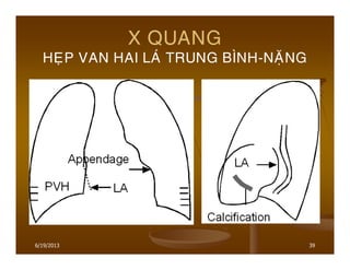
- 39. 6/19/2013 39 X QUANG HE├ÅP VAN HAI LA├Ö TRUNG B├īNH-NA├ŗNG
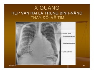
- 40. 6/19/2013 40 X QUANG HE├ÅP VAN HAI LA├Ö TRUNG B├īNH-NA├ŗNG THAY ├æO├ģI VE├Ć TIM
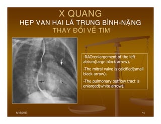
- 41. 6/19/2013 41 X QUANG HE├ÅP VAN HAI LA├Ö TRUNG B├īNH-NA├ŗNG THAY ├æO├ģI VE├Ć TIM -RAO:enlargement of the left atrium(large black arrow). -The mitral valve is calcified(small black arrow). -The pulmonary outflow tract is enlarged(white arrow).
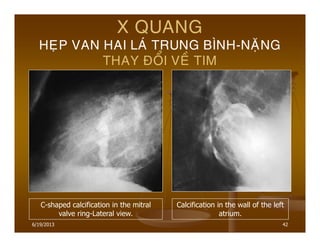
- 42. 6/19/2013 42 X QUANG HE├ÅP VAN HAI LA├Ö TRUNG B├īNH-NA├ŗNG THAY ├æO├ģI VE├Ć TIM C-shaped calcification in the mitral valve ring-Lateral view. Calcification in the wall of the left atrium.
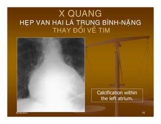
- 43. 6/19/2013 43 X QUANG HE├ÅP VAN HAI LA├Ö TRUNG B├īNH-NA├ŗNG THAY ├æO├ģI VE├Ć TIM Calcification within the left atrium.
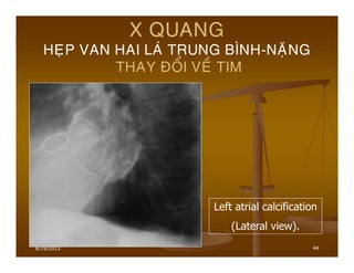
- 44. 6/19/2013 44 X QUANG HE├ÅP VAN HAI LA├Ö TRUNG B├īNH-NA├ŗNG THAY ├æO├ģI VE├Ć TIM Left atrial calcification (Lateral view).
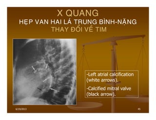
- 45. 6/19/2013 45 X QUANG HE├ÅP VAN HAI LA├Ö TRUNG B├īNH-NA├ŗNG THAY ├æO├ģI VE├Ć TIM -Left atrial calcification (white arrows). -Calcified mitral valve (black arrow).
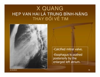
- 46. 6/19/2013 46 X QUANG HE├ÅP VAN HAI LA├Ö TRUNG B├īNH-NA├ŗNG THAY ├æO├ģI VE├Ć TIM -Calcified mitral valve. -Esophagus is pushed posteriorly by the enlarged left atrium.
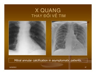
- 47. 6/19/2013 47 X QUANG THAY ├æO├ģI VE├Ć TIM Mitral annular calcification in asymptomatic patients.
- 48. 6/19/2013 48 X QUANG HE├ÅP VAN HAI LA├Ö TRUNG B├īNH-NA├ŗNG THAY ├æO├ģI VE├Ć TUA├ĆN HOA├śN PHO├ģI Ta├¬ng tua├Ān hoa├Ėn pho├źi thu├» ├▒o├żng (Ta├¬ng tua├Ān hoa├Ėn pho├źi sau mao ma├»ch): Ta├╣i pha├ón pho├Īi ma├»ch ma├╣u pho├źi xua├Īt hie├żn s├┤├╣m khi ALTT>12mmHg. Xua├Īt hie├żn ca├╣c ├▒├Č├┤├Ėng Kerley A,B,C,D khi ALTT>18mmHg.Th├Č├┤├Ėng tha├Īy nha├Īt la├Ė Kerley B.
- 49. 6/19/2013 49 X QUANG HE├ÅP VAN HAI LA├Ö TRUNG B├īNH-NA├ŗNG THAY ├æO├ģI VE├Ć TUA├ĆN HOA├śN PHO├ģI ├æMP to da├Ān Kho├╣ cha├źn ├▒oa├╣n pha├ón bie├żt gi├Č├Ąa CIA v├┤├╣i RM+HVD. CIA:Nh├│ (T) kho├óng to,kho├óng co├╣ ta├╣i pha├ón pho├Īi. Ve├Ā sau,├æMP to ├┤├╗ trung ta├óm,nho├╗ ├┤├╗ ngoa├»i vi (T├¼nh tra├»ng ta├¬ng a├╣p ├æMP la├óu nga├Ėy Tha├Ėnh tie├źu ├▒o├żng ma├»ch da├Ėy le├ón).
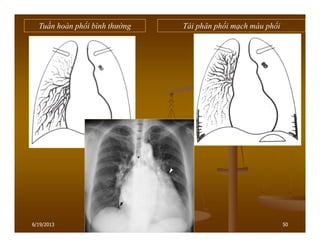
- 50. 50 Tuß║¦n ho├Ān phß╗Ģi b├¼nh thŲ░ß╗Øng T├Īi ph├ón phß╗æi mß║Īch m├Īu phß╗Ģi 6/19/2013
- 51. 516/19/2013
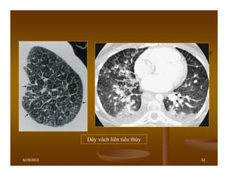
- 52. 52 D├Āy v├Īch li├¬n tiß╗āu th├╣y 6/19/2013
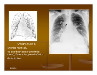
- 53. 53 CARDIAC FAILURE -Enlarged heart size. -No clear heart border (interstitiel edema), KerleyŌĆÖs line, pleural effusion. -Redistribution. 6/19/2013
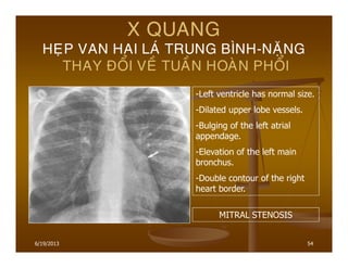
- 54. 6/19/2013 54 X QUANG HE├ÅP VAN HAI LA├Ö TRUNG B├īNH-NA├ŗNG THAY ├æO├ģI VE├Ć TUA├ĆN HOA├śN PHO├ģI -Left ventricle has normal size. -Dilated upper lobe vessels. -Bulging of the left atrial appendage. -Elevation of the left main bronchus. -Double contour of the right heart border. MITRAL STENOSIS
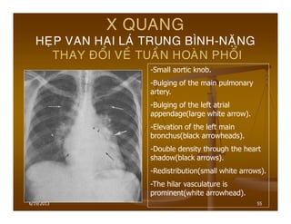
- 55. 6/19/2013 55 X QUANG HE├ÅP VAN HAI LA├Ö TRUNG B├īNH-NA├ŗNG THAY ├æO├ģI VE├Ć TUA├ĆN HOA├śN PHO├ģI -Small aortic knob. -Bulging of the main pulmonary artery. -Bulging of the left atrial appendage(large white arrow). -Elevation of the left main bronchus(black arrowheads). -Double density through the heart shadow(black arrows). -Redistribution(small white arrows). -The hilar vasculature is prominent(white arrowhead).
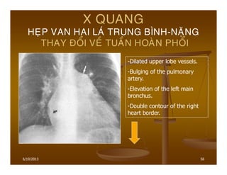
- 56. 6/19/2013 56 X QUANG HE├ÅP VAN HAI LA├Ö TRUNG B├īNH-NA├ŗNG THAY ├æO├ģI VE├Ć TUA├ĆN HOA├śN PHO├ģI -Dilated upper lobe vessels. -Bulging of the pulmonary artery. -Elevation of the left main bronchus. -Double contour of the right heart border.
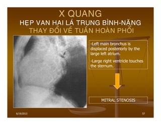
- 57. 6/19/2013 57 X QUANG HE├ÅP VAN HAI LA├Ö TRUNG B├īNH-NA├ŗNG THAY ├æO├ģI VE├Ć TUA├ĆN HOA├śN PHO├ģI -Left main bronchus is displaced posteriorly by the large left atrium. -Large right ventricle touches the sternum. MITRAL STENOSIS
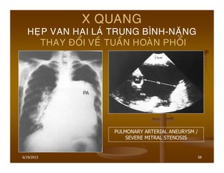
- 58. 6/19/2013 58 X QUANG HE├ÅP VAN HAI LA├Ö TRUNG B├īNH-NA├ŗNG THAY ├æO├ģI VE├Ć TUA├ĆN HOA├śN PHO├ģI PULMONARY ARTERIAL ANEURYSM / SEVERE MITRAL STENOSIS
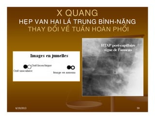
- 59. 6/19/2013 59 X QUANG HE├ÅP VAN HAI LA├Ö TRUNG B├īNH-NA├ŗNG THAY ├æO├ģI VE├Ć TUA├ĆN HOA├śN PHO├ģI
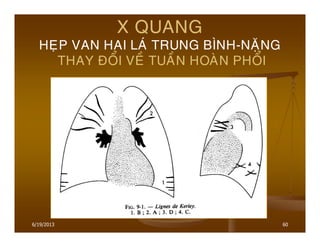
- 60. 6/19/2013 60 X QUANG HE├ÅP VAN HAI LA├Ö TRUNG B├īNH-NA├ŗNG THAY ├æO├ģI VE├Ć TUA├ĆN HOA├śN PHO├ģI
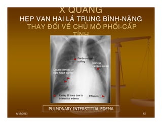
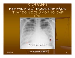
- 61. 6/19/2013 61 X QUANG HE├ÅP VAN HAI LA├Ö TRUNG B├īNH-NA├ŗNG THAY ├æO├ģI VE├Ć CHU├ø MO├é PHO├ģI-CA├üP T├ŹNH Phu├Ė pho├źi mo├ó ke├Ą. Phu├Ė pho├źi phe├Ī nang.
- 62. 6/19/2013 62 X QUANG HE├ÅP VAN HAI LA├Ö TRUNG B├īNH-NA├ŗNG THAY ├æO├ģI VE├Ć CHU├ø MO├é PHO├ģI-CA├üP T├ŹNH PULMONARY INTERSTITIAL EDEMA
- 63. 6/19/2013 63 X QUANG HE├ÅP VAN HAI LA├Ö TRUNG B├īNH-NA├ŗNG THAY ├æO├ģI VE├Ć CHU├ø MO├é PHO├ģI-CA├üP T├ŹNH PULMONARY EDEMA
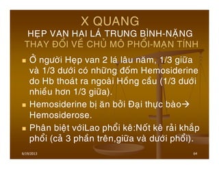
- 64. 6/19/2013 64 X QUANG HE├ÅP VAN HAI LA├Ö TRUNG B├īNH-NA├ŗNG THAY ├æO├ģI VE├Ć CHU├ø MO├é PHO├ģI-MA├ÅN T├ŹNH ├ö├ø ng├Č├┤├Ėi He├»p van 2 la├╣ la├óu na├¬m, 1/3 gi├Č├Ąa va├Ė 1/3 d├Č├┤├╣i co├╣ nh├Č├Ąng ├▒o├Īm Hemosiderine do Hb thoa├╣t ra ngoa├Ėi Ho├Āng ca├Āu (1/3 d├Č├┤├╣i nhie├Āu h├┤n 1/3 gi├Č├Ąa). Hemosiderine b├▓ a├¬n b├┤├╗i ├æa├»i th├Č├»c ba├Ėo Hemosiderose. Pha├ón bie├żt v├┤├╣iLao pho├źi ke├ó:No├Īt ke├ó ra├╗i kha├®p pho├źi (ca├╗ 3 pha├Ān tre├ón,gi├Č├Ąa va├Ė d├Č├┤├╣i pho├źi).
- 65. 6/19/2013 65 X QUANG HE├ÅP VAN HAI LA├Ö TRUNG B├īNH-NA├ŗNG THAY ├æO├ģI VE├Ć CHU├ø MO├é PHO├ģI-MA├ÅN T├ŹNH X├┤ pho├źi:nh├Č├Ąng da├╗i m├┤├Ė sa├®c ne├╣t. Da├Ėy d├Łnh ma├Ėng pho├źi:do thoa├╣t d├▓ch Da├Ėy d├Łnh. Nh├Č├Ąng vu├Ėng m├┤├Ė cu├╗a nho├Āi ma├╣u pho├źi cu├Ą.
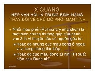
- 66. 6/19/2013 66 X QUANG HE├ÅP VAN HAI LA├Ö TRUNG B├īNH-NA├ŗNG THAY ├æO├ģI VE├Ć CHU├ø MO├é PHO├ģI-MA├ÅN T├ŹNH Nho├Āi ma├╣u pho├źi (Pulmonary infarction) la├Ė mo├żt bie├Īn ch├Č├╣ng th├Č├┤├Ėng ga├½p cu├╗a be├żnh van 2 la├╣ v├¼ thuye├ón ta├®c co├╣ nguo├Ān go├Īc t├Č├Ė: Hoa├½c do nh├Č├Ąng cu├»c ma├╣u ├▒o├óng ├┤├╗ ngoa├»i vi v├¼ cung l├Č├┤├»ng tim tha├Īp. Hoa├½c do cu├»c ma├╣u ├▒o├óng t├Č├Ė Nh├│ (P) xua├Īt hie├żn sau Rung nh├│.
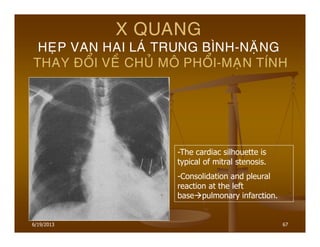
- 67. 6/19/2013 67 X QUANG HE├ÅP VAN HAI LA├Ö TRUNG B├īNH-NA├ŗNG THAY ├æO├ģI VE├Ć CHU├ø MO├é PHO├ģI-MA├ÅN T├ŹNH -The cardiac silhouette is typical of mitral stenosis. -Consolidation and pleural reaction at the left base pulmonary infarction.
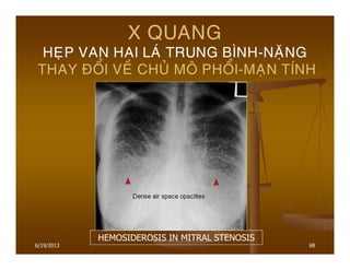
- 68. 6/19/2013 68 X QUANG HE├ÅP VAN HAI LA├Ö TRUNG B├īNH-NA├ŗNG THAY ├æO├ģI VE├Ć CHU├ø MO├é PHO├ģI-MA├ÅN T├ŹNH HEMOSIDEROSIS IN MITRAL STENOSIS
- 69. 6/19/2013 69 X QUANG HE├ÅP VAN HAI LA├Ö TRUNG B├īNH-NA├ŗNG THAY ├æO├ģI VE├Ć CHU├ø MO├é PHO├ģI-MA├ÅN T├ŹNH KERLEYŌĆÖS B LINES & HEMOSIDEROSIS IN MITRAL STENOSIS
- 70. 6/19/2013 70 ├æA├ÅI C├¢├öNG H├ö├ø VAN HAI LA├Ö (MITRAL REGURGITATION) H├┤├╗ van 2 la├╣ co├╣ the├ź ca├Īp t├Łnh:├æ├Č├╣t da├óy cha├©ng-co├żt c├┤ (sau Nho├Āi ma├╣u c├┤ tim,Vie├óm no├żi ta├óm ma├»c nhie├Żm tru├Ėng) H├┤├╗ 2 la├╣ nh├Čng Tha├Īt (T) ch├Ča k├▓p gia├Ąn ro├żng. H├┤├╗ van 2 la├╣ co├╣ the├ź ma├»n t├Łnh:Sau RAA, hoa├½c do lo├Ż van gia├Ąn ro├żng (Ho├żi ch├Č├╣ng Marfan, Be├żnh c├┤ tim)ŌĆ”
- 71. 6/19/2013 71 X QUANG H├ö├ø VAN HAI LA├Ö NHE├Å Tha├Īt (T) va├Ė Nh├│ (T) gia├Ąn l├┤├╣n do ŌĆ£overloadŌĆØ. Nh├│ (T) co├Ėn bu├Ė X quang tim-pho├źi b├¼nh th├Č├┤├Ėng du├Ė tre├ón La├óm sa├Ėng nghe ATTT. Do va├ży,cu├Ąng nh├Č He├»p van 2 la├╣,X quang tim-pho├źi b├¼nh th├Č├┤├Ėng kho├óng loa├»i tr├Č├Ė ├▒├Č├┤├»c H├┤├╗ van 2 la├╣.
- 72. 6/19/2013 72 X QUANG H├ö├ø VAN HAI LA├Ö TRUNG B├īNH Nh├│ (T) co├Ėn bu├Ė. Nh├│ (T) to ra,nh├Čng a├╣p sua├Īt ch├Ča ta├¬ng nhie├Āu. Tha├Īt (T) cu├Ąng gia├Ąn to v├Č├Ėa pha├╗i. Quai ├æMC nho├╗ do cung l├Č├┤├»ng tim gia├╗m.
- 73. 6/19/2013 73 X QUANG H├ö├ø VAN HAI LA├Ö NA├ŗNG Nh├│ (T) ma├Īt bu├Ė. Nh├│ (T) to ra,a├╣p sua├Īt ta├¬ng cao.Ne├Īu so sa├╣nh v├┤├╣i He├»p 2 la├╣ ,th├¼ Nh├│ (T) to h├┤n nhie├Āu,co├╣ khi v├Č├┤├»t ra ngoa├Ėi b├┤├Ė cu├╗a Nh├│ (P). Tha├Īt (T) to ra,ta├»o ne├ón h├¼nh a├╗nh ŌĆ£big heart diseaseŌĆØ. Tre├ón h├¼nh nghie├óng hoa├½c LAO: Tha├Īt (T) to che la├Īp khoa├╗ng sa├╣ng sau tim.
- 74. 6/19/2013 74 X QUANG H├ö├ø VAN HAI LA├Ö NA├ŗNG Quai ├æMC nho├╗ do cung l├Č├┤├»ng tim gia├╗m. Ph├¼nh cung ├æMP ŌĆØMitralisation du bord gauche du coeurŌĆØ. Ta├╣i pha├ón pho├Īi tua├Ān hoa├Ėn pho├źi. Ca├╣c ├▒├Č├┤├Ėng Kerley ├Łt ga├½p h├┤n va├Ė kho├óng ro├Ą so v├┤├╣i He├»p van 2 la├╣.
- 75. 6/19/2013 75 X QUANG H├ö├ø VAN HAI LA├Ö NA├ŗNG MITRAL REGURGITATION
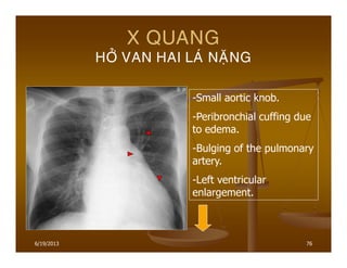
- 76. 6/19/2013 76 X QUANG H├ö├ø VAN HAI LA├Ö NA├ŗNG -Small aortic knob. -Peribronchial cuffing due to edema. -Bulging of the pulmonary artery. -Left ventricular enlargement.
- 77. 6/19/2013 77 X QUANG H├ö├ø VAN HAI LA├Ö NA├ŗNG MITRAL REGURGITATION
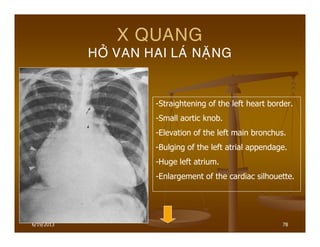
- 78. 6/19/2013 78 X QUANG H├ö├ø VAN HAI LA├Ö NA├ŗNG -Straightening of the left heart border. -Small aortic knob. -Elevation of the left main bronchus. -Bulging of the left atrial appendage. -Huge left atrium. -Enlargement of the cardiac silhouette.
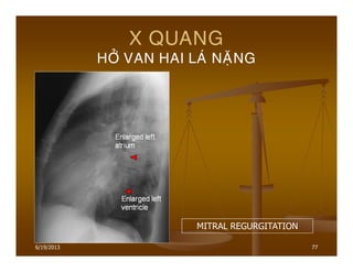
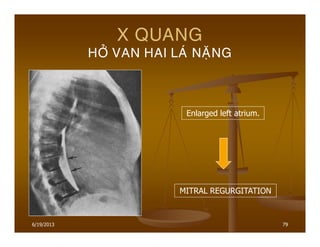
- 79. 6/19/2013 79 X QUANG H├ö├ø VAN HAI LA├Ö NA├ŗNG Enlarged left atrium. MITRAL REGURGITATION
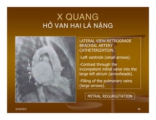
- 80. 6/19/2013 80 X QUANG H├ö├ø VAN HAI LA├Ö NA├ŗNG LATERAL VIEW:RETROGRADE BRACHIAL ARTERY CATHETERIZATION. -Left ventricle (small arrows). -Contrast through the incompetent mitral valve into the large left atrium (arrowheads). -Filling of the pulmonary veins (large arrows). MITRAL REGURGITATION
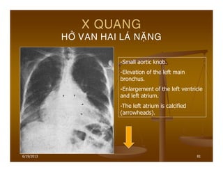
- 81. 6/19/2013 81 X QUANG H├ö├ø VAN HAI LA├Ö NA├ŗNG -Small aortic knob. -Elevation of the left main bronchus. -Enlargement of the left ventricle and left atrium. -The left atrium is calcified (arrowheads).
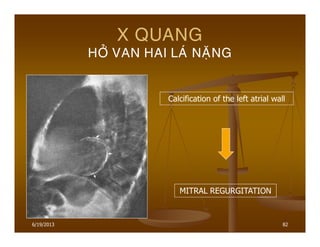
- 82. 6/19/2013 82 X QUANG H├ö├ø VAN HAI LA├Ö NA├ŗNG Calcification of the left atrial wall MITRAL REGURGITATION
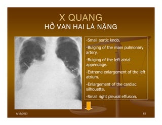
- 83. 6/19/2013 83 X QUANG H├ö├ø VAN HAI LA├Ö NA├ŗNG -Small aortic knob. -Bulging of the main pulmonary artery. -Bulging of the left atrial appendage. -Extreme enlargement of the left atrium. -Enlargement of the cardiac silhouette. -Small right pleural effusion.
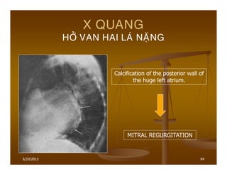
- 84. 6/19/2013 84 X QUANG H├ö├ø VAN HAI LA├Ö NA├ŗNG Calcification of the posterior wall of the huge left atrium. MITRAL REGURGITATION
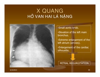
- 85. 6/19/2013 85 X QUANG H├ö├ø VAN HAI LA├Ö NA├ŗNG MITRAL REGURGITATION -Small aortic knob. -Elevation of the left main bronchus. -Extreme enlargement of the left atrium (arrows). -Enlargement of the cardiac silhouette.
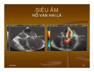
- 86. 6/19/2013 86 SIE├éU A├éM H├ö├ø VAN HAI LA├Ö
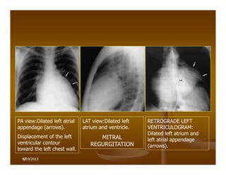
- 87. 87 PA view:Dilated left atrial appendage (arrows). Displacement of the left ventricular contour toward the left chest wall. LAT view:Dilated left atrium and ventricle. MITRAL REGURGITATION RETROGRADE LEFT VENTRICULOGRAM: Dilated left atrium and left atrial appendage (arrows). 6/19/2013
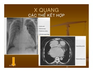
- 88. 6/19/2013 88 X QUANG CA├ÖC THE├ģ KE├üT H├ö├ÅP He├»p+H├┤├╗ van 2 la├╣:Nh├│ (T) ra├Īt l├┤├╣n,co├╣ ├▒o├╣ng vo├ói ├┤├╗ van 2 la├╣ va├Ė tha├Ėnh Nh├│ (T),tim to toa├Ėn bo├ż. H├┤├╗ van 2 la├╣+Be├żnh van ├æMC(He├»p,h├┤├╗). H├┤├╗ van 2 la├╣+H├┤├╗ van 3 la├╣. H├┤├╗ van 2 la├╣+H├┤├╗ van 3 la├╣+Be├żnh van ├æMC.
- 89. 6/19/2013 89 X QUANG CA├ÖC THE├ģ KE├üT H├ö├ÅP
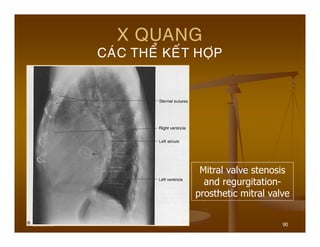
- 90. 6/19/2013 90 X QUANG CA├ÖC THE├ģ KE├üT H├ö├ÅP Mitral valve stenosis and regurgitation- prosthetic mitral valve
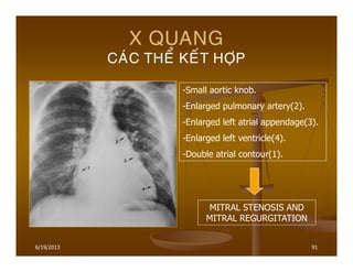
- 91. 6/19/2013 91 X QUANG CA├ÖC THE├ģ KE├üT H├ö├ÅP MITRAL STENOSIS AND MITRAL REGURGITATION -Small aortic knob. -Enlarged pulmonary artery(2). -Enlarged left atrial appendage(3). -Enlarged left ventricle(4). -Double atrial contour(1).
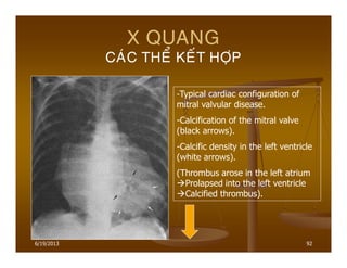
- 92. 6/19/2013 92 X QUANG CA├ÖC THE├ģ KE├üT H├ö├ÅP -Typical cardiac configuration of mitral valvular disease. -Calcification of the mitral valve (black arrows). -Calcific density in the left ventricle (white arrows). (Thrombus arose in the left atrium Prolapsed into the left ventricle Calcified thrombus).
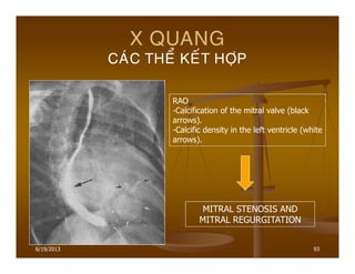
- 93. 6/19/2013 93 X QUANG CA├ÖC THE├ģ KE├üT H├ö├ÅP RAO -Calcification of the mitral valve (black arrows). -Calcific density in the left ventricle (white arrows). MITRAL STENOSIS AND MITRAL REGURGITATION
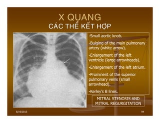
- 94. 6/19/2013 94 X QUANG CA├ÖC THE├ģ KE├üT H├ö├ÅP -Small aortic knob. -Bulging of the main pulmonary artery (white arrow). -Enlargement of the left ventricle (large arrowheads). -Enlargement of the left atrium. -Prominent of the superior pulmonary veins (small arrowhead). -KerleyŌĆÖs B lines. MITRAL STENOSIS AND MITRAL REGURGITATION
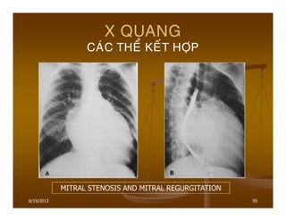
- 95. 6/19/2013 95 X QUANG CA├ÖC THE├ģ KE├üT H├ö├ÅP MITRAL STENOSIS AND MITRAL REGURGITATION
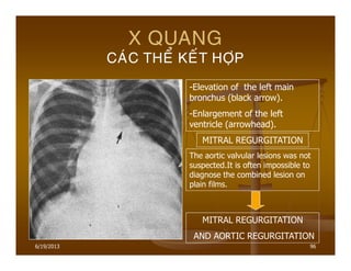
- 96. 6/19/2013 96 X QUANG CA├ÖC THE├ģ KE├üT H├ö├ÅP -Elevation of the left main bronchus (black arrow). -Enlargement of the left ventricle (arrowhead). MITRAL REGURGITATION The aortic valvular lesions was not suspected.It is often impossible to diagnose the combined lesion on plain films. MITRAL REGURGITATION AND AORTIC REGURGITATION
- 97. 6/19/2013 97 X QUANG HE├ÅP VAN HAI LA├Ö TRUNG B├īNH-NA├ŗNG THAY ├æO├ģI VE├Ć TIM SEVERE MITRAL VALVE DISEASE
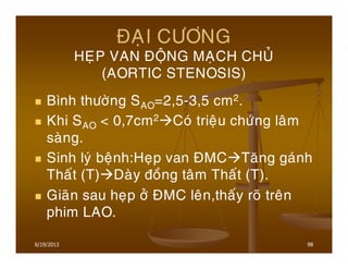
- 98. 6/19/2013 98 ├æA├ÅI C├¢├öNG HE├ÅP VAN ├æO├äNG MA├ÅCH CHU├ø (AORTIC STENOSIS) B├¼nh th├Č├┤├Ėng SAO=2,5-3,5 cm2. Khi SAO < 0,7cm2 Co├╣ trie├żu ch├Č├╣ng la├óm sa├Ėng. Sinh ly├╣ be├żnh:He├»p van ├æMC Ta├¬ng ga├╣nh Tha├Īt (T) Da├Ėy ├▒o├Āng ta├óm Tha├Īt (T). Gia├Ąn sau he├»p ├┤├╗ ├æMC le├ón,tha├Īy ro├Ą tre├ón phim LAO.
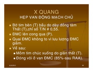
- 99. 6/19/2013 99 X QUANG HE├ÅP VAN ├æO├äNG MA├ÅCH CHU├ø B├┤├Ė tim be├ón (T) ba├Āu do da├Ėy ├▒o├Āng ta├óm Tha├Īt (T),ch├” so├Ī T/N # 0,55. ├æMC le├ón cong qua (P). Quai ├æMC kho├óng to v├¼ l├Ču l├Č├┤├»ng ├æMC gia├╗m. Ve├Ā sau: Mo├╗m tim chu├╣c xuo├Īng do gia├Ąn tha├Īt (T). ├æo├╣ng vo├ói ├┤├╗ van ├æMC (85%-sau RAA).
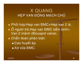
- 100. 6/19/2013 100 X QUANG HE├ÅP VAN ├æO├äNG MA├ÅCH CHU├ø Pho├Īi h├┤├»p:He├»p van ├æMC+He├»p van 2 la├╣. ├ö├ø ng├Č├┤├Ėi tre├╗,He├»p van ├æMC ba├źm sinh= Van 2 ma├╗nh (Bicuspid valve). Cha├źn ├▒oa├╣n pha├ón bie├żt: Cao huye├Īt a├╣p. X├┤ v├Č├Ąa ├æMC.
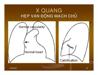
- 101. 6/19/2013 101 X QUANG HE├ÅP VAN ├æO├äNG MA├ÅCH CHU├ø
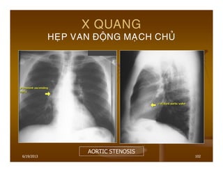
- 102. 6/19/2013 102 X QUANG HE├ÅP VAN ├æO├äNG MA├ÅCH CHU├ø AORTIC STENOSIS
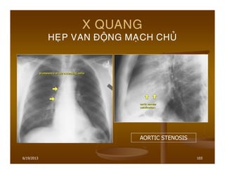
- 103. 6/19/2013 103 X QUANG HE├ÅP VAN ├æO├äNG MA├ÅCH CHU├ø AORTIC STENOSIS
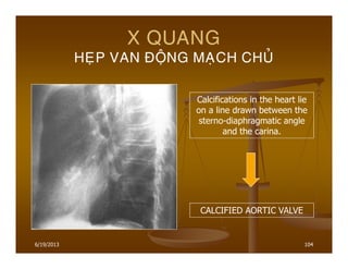
- 104. 6/19/2013 104 X QUANG HE├ÅP VAN ├æO├äNG MA├ÅCH CHU├ø Calcifications in the heart lie on a line drawn between the sterno-diaphragmatic angle and the carina. CALCIFIED AORTIC VALVE
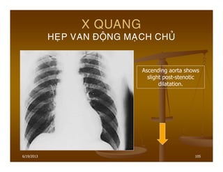
- 105. 6/19/2013 105 X QUANG HE├ÅP VAN ├æO├äNG MA├ÅCH CHU├ø Ascending aorta shows slight post-stenotic dilatation.
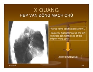
- 106. 6/19/2013 106 X QUANG HE├ÅP VAN ├æO├äNG MA├ÅCH CHU├ø -Aortic valve calcification (arrow). -Posterior displacement of the left ventricle behind the line of the inferior vena cava. AORTIC STENOSIS
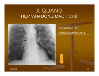
- 107. 6/19/2013 107 X QUANG HE├ÅP VAN ├æO├äNG MA├ÅCH CHU├ø -Normal heart size. -Dilated ascending aorta.
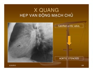
- 108. 6/19/2013 108 X QUANG HE├ÅP VAN ├æO├äNG MA├ÅCH CHU├ø Calcified aortic valve. AORTIC STENOSIS
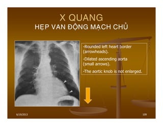
- 109. 6/19/2013 109 X QUANG HE├ÅP VAN ├æO├äNG MA├ÅCH CHU├ø -Rounded left heart border (arrowheads). -Dilated ascending aorta (small arrows). -The aortic knob is not enlarged.
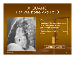
- 110. 6/19/2013 110 X QUANG HE├ÅP VAN ├æO├äNG MA├ÅCH CHU├ø LAO -Bulging of the ascending aorta because of post-stenotic dilatation (arrowheads). -Calcified aortic valve (black arrow). AORTIC STENOSIS
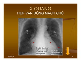
- 111. 6/19/2013 111 X QUANG HE├ÅP VAN ├æO├äNG MA├ÅCH CHU├ø
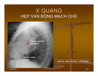
- 112. 6/19/2013 112 X QUANG HE├ÅP VAN ├æO├äNG MA├ÅCH CHU├ø AORTIC AND MITRAL STENOSIS
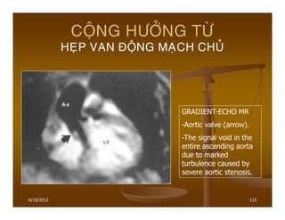
- 113. 6/19/2013 113 CO├äNG H├¢├ö├øNG T├¢├ś HE├ÅP VAN ├æO├äNG MA├ÅCH CHU├ø GRADIENT-ECHO MR -Aortic valve (arrow). -The signal void in the entire ascending aorta due to marked turbulence caused by severe aortic stenosis.
- 114. 6/19/2013 114 X QUANG H├ö├ø VAN ├æO├äNG MA├ÅCH CHU├ø Nguye├ón nha├ón th├Č├┤├Ėng ga├½p nha├Īt la├Ė sau RAA. Ca├╣c nguye├ón nha├ón kha├╣c bao go├Ām:Giang mai,Vie├óm no├żi ta├óm ma├»c nhie├Żm tru├Ėng,Ho├żi ch├Č├╣ng Marfan,Cha├Īn th├Č├┤ng,├æMC bo├╣c ta├╣ch,Vie├óm kh├┤├╣p da├»ng tha├Īp,Thoa├╣i ho├╣a sau ghe├╣p van sinh ho├»c.
- 115. 6/19/2013 115 X QUANG H├ö├ø VAN ├æO├äNG MA├ÅCH CHU├ø Sinh ly├╣ be├żnh:Ma├╣u phu├»t ng├Č├┤├»c va├Ėo Tha├Īt (T) trong ky├Ė ta├óm tr├Č├┤ng Gia├Ąn buo├Āng tha├Īt (T).Tha├Īt (T) gia├Ąn Gia├Ąn vo├Ėng van 2 la├╣ H├┤├╗ van 2 la├╣ Gia├Ąn buo├Āng Nh├│ (T). Mo├╗m tim chu├╣c xuo├Īng d├Č├┤├╣i va├Ė ra sau B├┤├Ė (T) tim thoai thoa├╗i va├Ė ├▒├”nh tim ├┤├╗ d├Č├┤├╣i vo├Ėm hoa├Ėnh (T). Ch├” so├Ī T/N > 0,6.
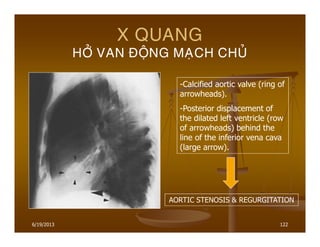
- 116. 6/19/2013 116 X QUANG H├ö├ø VAN ├æO├äNG MA├ÅCH CHU├ø ├æMP kho├óng to,co├╣ khi nh├Č lo├Ąm va├Ėo. ├æMC le├ón th├Č├┤├Ėng kho├óng gia├Ąn,ne├Īu ├æMC le├ón gia├Ąn ro├Ą,pha├╗i coi ch├Č├Ėng co├╣ ke├Īt h├┤├»p v├┤├╣i He├»p van ├æMC. Quai ├æMC ha├Āu nh├Č kho├óng thay ├▒o├źi. Chie├Īu X quang:├æMC ├▒a├żp ma├»nh.
- 117. 6/19/2013 117 X QUANG H├ö├ø VAN ├æO├äNG MA├ÅCH CHU├ø
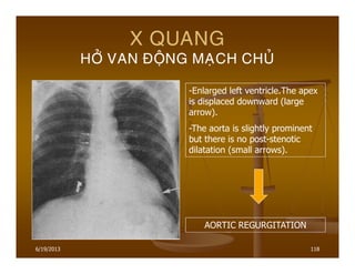
- 118. 6/19/2013 118 X QUANG H├ö├ø VAN ├æO├äNG MA├ÅCH CHU├ø -Enlarged left ventricle.The apex is displaced downward (large arrow). -The aorta is slightly prominent but there is no post-stenotic dilatation (small arrows). AORTIC REGURGITATION
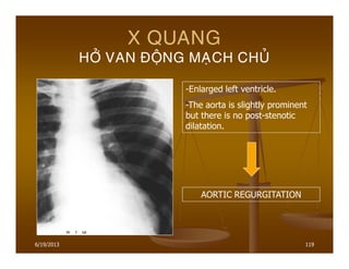
- 119. 6/19/2013 119 X QUANG H├ö├ø VAN ├æO├äNG MA├ÅCH CHU├ø -Enlarged left ventricle. -The aorta is slightly prominent but there is no post-stenotic dilatation. AORTIC REGURGITATION
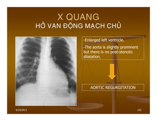
- 120. 6/19/2013 120 X QUANG H├ö├ø VAN ├æO├äNG MA├ÅCH CHU├ø -Enlarged left ventricle. -The aorta is slightly prominent but there is no post-stenotic dilatation. AORTIC REGURGITATION
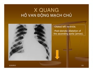
- 121. 6/19/2013 121 X QUANG H├ö├ø VAN ├æO├äNG MA├ÅCH CHU├ø -Dilated left ventricle. -Post-stenotic dilatation of the ascending aorta (arrow).
- 122. 6/19/2013 122 X QUANG H├ö├ø VAN ├æO├äNG MA├ÅCH CHU├ø -Calcified aortic valve (ring of arrowheads). -Posterior displacement of the dilated left ventricle (row of arrowheads) behind the line of the inferior vena cava (large arrow). AORTIC STENOSIS & REGURGITATION
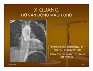
- 123. 6/19/2013 123 X QUANG H├ö├ø VAN ├æO├äNG MA├ÅCH CHU├ø RETROGRADE AORTOGRAM IN AORTIC REGURGITATION Reflux flow (arrows) in the dilated left ventricle.
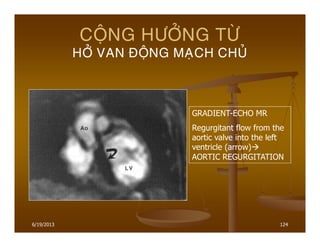
- 124. 6/19/2013 124 CO├äNG H├¢├ö├øNG T├¢├ś H├ö├ø VAN ├æO├äNG MA├ÅCH CHU├ø GRADIENT-ECHO MR Regurgitant flow from the aortic valve into the left ventricle (arrow) AORTIC REGURGITATION
- 125. 6/19/2013 125 ├æA├ÅI C├¢├öNG BE├äNH VAN ├æO├äNG MA├ÅCH PHO├ģI MA├ēC PHA├øI (ACQUIRED PULMONARY VALVULAR LESIONS) Be├żnh hie├Īm khi ├▒├┤n ├▒o├żc va├Ė hie├Īm khi na├½ng. Be├żnh co├╣ the├ź tha├Īy trong Ho├żi ch├Č├╣ng carcinoid. H├¼nh a├╗nh X quang th├Č├┤├Ėng ke├Īt h├┤├»p to├źn th├Č├┤ng ca├╣c van tim kha├╣c.
- 126. 6/19/2013 126 ├æA├ÅI C├¢├öNG BE├äNH VAN BA LA├Ö MA├ēC PHA├øI (ACQUIRED TRICUSPID LESIONS) T├Č├┤ng ├▒o├Īi ga├½p nhie├Āu h├┤n be├żnh van ├æMP ma├®c pha├╗i. Be├żnh th├Č├┤├Ėng ga├½p nha├Īt sau RAA(Be├żnh ha├Āu nh├Č luo├ón luo├ón ke├Īt h├┤├»p v├┤├╣i be├żnh ly├╣ van 2 la├╣ va├Ė be├żnh ly├╣ van ├æMC). Ca├╣c nguye├ón nha├ón kha├╣c:Vie├óm no├żi ta├óm ma├»c nhie├Żm tru├Ėng,Cha├Īn th├Č├┤ng ng├Č├»c,B├Č├┤├╣u carcinoidŌĆ”
- 127. 6/19/2013 127 X QUANG BE├äNH VAN BA LA├Ö MA├ēC PHA├øI Gia├Ąn buo├Āng Tha├Īt (P) va├Ė Nh├│ (P). Th├Č├┤├Ėng kho├óng vo├ói ho├╣a van 3 la├╣. Gia├Ąn TMC tre├ón,TM azygos. Da├Īu hie├żu phu├»:Gan to,├▒o├żi vo├Ėm hoa├Ėnh (P) le├ón cao. Khi H├┤├╗ van 3 la├╣+He├»p van 2 la├╣ Da├Īu ta├╣i pha├ón pho├Īi va├Ė ├▒├Č├┤├Ėng Kerley ga├Ān nh├Č bie├Īn ma├Īt.
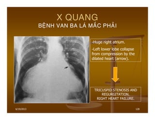
- 128. 6/19/2013 128 X QUANG BE├äNH VAN BA LA├Ö MA├ēC PHA├øI -Huge right atrium. -Left lower lobe collapse from compression by the dilated heart (arrow). TRICUSPID STENOSIS AND REGURGITATION. RIGHT HEART FAILURE.
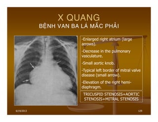
- 129. 6/19/2013 129 X QUANG BE├äNH VAN BA LA├Ö MA├ēC PHA├øI -Enlarged right atrium (large arrows). -Decrease in the pulmonary vasculature. -Small aortic knob. -Typical left border of mitral valve disease (small arrow). -Elevation of the right hemi- diaphragm. TRICUSPID STENOSIS+AORTIC STENOSIS+MITRAL STENOSIS
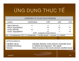
- 130. 6/19/2013 130 ÛÙNG DUÏNG THÛÏC TEÁ
- 131. 6/19/2013 131 KE├üT LUA├äN Be├żnh van tim ma├®c pha├╗i pha├Ān l├┤├╣n do RAA. To├źn th├Č├┤ng th├Č├┤├Ėng ga├½p nha├Īt ├┤├╗ van 2 la├╣, tie├Īp ├▒e├Īn la├Ė van ├æMC. Co├╣ the├ź ch├” He├»p ├▒├┤n thua├Ān,H├┤├╗ ├▒├┤n thua├Ān, co├╣ the├ź He├»p-H├┤├╗ ke├Īt h├┤├»p,co├╣ the├ź to├źn th├Č├┤ng nhie├Āu van cu├Ėng mo├żt lu├╣c.
- 132. 6/19/2013 132